Ki-67 Monoclonal Antibody (SolA15), Biotin, eBioscience™, Invitrogen™
Manufacturer: Fischer Scientific
Select a Size
| Pack Size | SKU | Availability | Price |
|---|---|---|---|
| Each of 1 | 50-112-2553-Each-of-1 | In Stock | ₹ 32,930.00 |
50-112-2553 - Each of 1
In Stock
Quantity
1
Base Price: ₹ 32,930.00
GST (18%): ₹ 5,927.40
Total Price: ₹ 38,857.40
Antigen
Ki-67
Classification
Monoclonal
Concentration
0.5 mg/mL
Formulation
PBS with 0.09% sodium azide; pH 7.2
Gene Accession No.
E9PVX6, P46013
Gene Symbols
Mki67
Purification Method
Affinity chromatography
Regulatory Status
RUO
Gene ID (Entrez)
100686578, 102135895, 17345, 291234, 4288
Content And Storage
4° C, store in dark, DO NOT FREEZE!
Form
Liquid
Applications
Flow Cytometry, Immunocytochemistry, Immunohistochemistry (Frozen), Immunohistochemistry (Paraffin)
Clone
SolA15
Conjugate
Biotin
Gene
Mki67
Gene Alias
antigen identified by monoclonal antibody Ki 67; antigen identified by monoclonal antibody Ki-67; Antigen identified by monoclonal antibody Ki-67 homolog; Antigen KI-67; Antigen KI-67 homolog; antigen KI-67; proliferation marker protein Ki-67; antigen KI-67-like; cb31; D630048A14Rik; I79_022666; Ki67; Ki-67; KIA; LOW QUALITY PROTEIN: proliferation marker protein Ki-67; marker of proliferation Ki-67; MIB-; MIB-1; Mki67; PPP1R105; Proliferation marker protein Ki-67; proliferation-related Ki-67 antigen; protein phosphatase 1, regulatory subunit 105; RP11-380J17.2; sb:cb31; si:ch211-250b22.7; unnamed protein product; wu:fa11g09; wu:fb57a07; wu:fi14e05
Host Species
Rat
Quantity
100 μg
Primary or Secondary
Primary
Target Species
Canine, Cynomolgus Monkey, Human, Mouse, Non-human Primate, Rat
Product Type
Antibody
Isotype
IgG2a κ
Description
- Description: The monoclonal antibody SolA15 recognizes mouse and rat Ki-67, a 300 kDa nuclear protein
- Ki-67 is present during all active phases of the cell cycle (G1, S, G2, and mitosis), but is absent from resting cells (G0)
- Ki-67 is detected within the nucleus during interphase but redistributes to the chromosomes during mitosis
- Ki-67 is used as a marker for determining the growth fraction of a given population of cells
- In studies of tumor cells, the Ki-67 labeling index refers to the number of Ki-67 positive cells within the population and this is used to predict outcome of particular cancer types
- Ki-67 has been shown to interact with the DNA-bound protein chromobox protein homolog 3 (CBX3) (heterochromatin)
- The SolA15 antibody also recognizes human, non-human primate and canine Ki-67
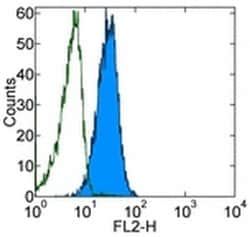
- Applications Reported: This SolA15 antibody has been reported for use in intracellular staining followed by flow cytometric analysis, immunohistochemical staining of frozen tissue sections, immunohistochemical staining of formalin-fixed paraffin embedded tissue sections, microscopy, and immunocytochemistry
- Applications Tested: This SolA15 antibody has been tested immunocytochemistry of fixed and permeabilized C2C12 cells and can be used at less than or equal to 5 μg/mL or intracellular staining and flow cytometric analysis of stimulated mous esplenocytes cells using the Foxp3/Transcription Factor Buffer Set (cat
- 00-5523) and protocol
- Ki-67 is a nuclear protein that is expressed during various stages in the cell cycle, particularly during late G1, S, G2, and M phases
- The protein has a forkhead associated domain (FHA) through which it associates with euchromatin at the perichromosomal layer, the centromeric heterochromatin, and the nucleolus
- Ki-67 is shown to have a cell cycle dependent topographical distribution with perinucleolar expression at G1, expression in the nuclear matrix at G2, and expression on the chromosomes during M phase
- Ki-67 is commonly used as a proliferation marker because it is not detected in G0 cells, but increases steadily from G1 through mitosis
- Ki-67 antibodies are useful in establishing the cell growing fraction in neoplasms
- In neoplastic tissues, the prognostic value is comparable to the tritiated thymidine-labelling index
- The correlation between low Ki-67 index and histologically low-grade tumors is strong
- Ki-67 is routinely used as a neuronal marker of cell cycling and proliferation.
Compare Similar Items
Show Difference

Antigen: Ki-67
Classification: Monoclonal
Concentration: 0.5 mg/mL
Formulation: PBS with 0.09% sodium azide; pH 7.2
Gene Accession No.: E9PVX6, P46013
Gene Symbols: Mki67
Purification Method: Affinity chromatography
Regulatory Status: RUO
Gene ID (Entrez): 100686578, 102135895, 17345, 291234, 4288
Content And Storage: 4° C, store in dark, DO NOT FREEZE!
Form: Liquid
Applications: Flow Cytometry, Immunocytochemistry, Immunohistochemistry (Frozen), Immunohistochemistry (Paraffin)
Clone: SolA15
Conjugate: Biotin
Gene: Mki67
Gene Alias: antigen identified by monoclonal antibody Ki 67; antigen identified by monoclonal antibody Ki-67; Antigen identified by monoclonal antibody Ki-67 homolog; Antigen KI-67; Antigen KI-67 homolog; antigen KI-67; proliferation marker protein Ki-67; antigen KI-67-like; cb31; D630048A14Rik; I79_022666; Ki67; Ki-67; KIA; LOW QUALITY PROTEIN: proliferation marker protein Ki-67; marker of proliferation Ki-67; MIB-; MIB-1; Mki67; PPP1R105; Proliferation marker protein Ki-67; proliferation-related Ki-67 antigen; protein phosphatase 1, regulatory subunit 105; RP11-380J17.2; sb:cb31; si:ch211-250b22.7; unnamed protein product; wu:fa11g09; wu:fb57a07; wu:fi14e05
Host Species: Rat
Quantity: 100 μg
Primary or Secondary: Primary
Target Species: Canine, Cynomolgus Monkey, Human, Mouse, Non-human Primate, Rat
Product Type: Antibody
Isotype: IgG2a κ

Antigen: OVA257-264 (SIINFEKL) peptide bound to H-2Kb
Classification: Monoclonal
Concentration: 0.5 mg/mL
Formulation: PBS with 0.09% sodium azide; pH 7.2
Gene Accession No.: __
Gene Symbols: __
Purification Method: Affinity chromatography
Regulatory Status: RUO
Gene ID (Entrez): __
Content And Storage: 4° C, store in dark, DO NOT FREEZE!
Form: Liquid
Applications: Flow Cytometry, Immunohistochemistry
Clone: eBio25-D1.16 (25-D1.16)
Conjugate: Biotin
Gene: __
Gene Alias: H-2Kb-SIINFEKL; OVA257-264; OVA257-264 H-2Kb; OVA-Kb
Host Species: Mouse
Quantity: 50 μg
Primary or Secondary: Primary
Target Species: Mouse
Product Type: Antibody
Isotype: IgG1 κ
Antigen: CD178 (Fas Ligand)
Classification: Monoclonal
Concentration: 0.5 mg/mL
Formulation: PBS with 0.09% sodium azide; pH 7.2
Gene Accession No.: P41047
Gene Symbols: Fasl
Purification Method: Affinity chromatography
Regulatory Status: RUO
Gene ID (Entrez): 14103
Content And Storage: 4° C, store in dark, DO NOT FREEZE!
Form: Liquid
Applications: Flow Cytometry
Clone: MFL3
Conjugate: Biotin
Gene: Fasl
Gene Alias: ADAM10-processed FasL form; ALPS1B; APL; Apoptosis (APO 1) antigen ligand 1; apoptosis (APO-1) antigen ligand 1; Apoptosis (APO-1) antigen ligand 1 (Fas antigen ligand); apoptosis antigen ligand; APT1LG1; APTL; CD178; CD178 antigen; CD95 ligand; CD95 ligand protein;Generalized lymphoproliferative disease (Gld); Cd95l; CD95-L; CD95L protein; fas antigen ligand; Fas ligand; Fas ligand (TNF superfamily, member 6); Fasl; Fas-L; FasL ICD; FasL intracellular domain; Faslg; Fas-LG; generalized lymphoproliferative disease; gld; mutant tumor necrosis factor family member 6; Receptor-binding FasL ectodomain; sFasL; Soluble Fas ligand; soluble form; SPA; SPPL2A-processed FasL form; TNFL; Tnfsf6; TNLG1A; Tumor necrosis factor (ligand) superfamily member 6; Tumor necrosis factor (ligand) superfamily member 6 (apoptosis (APO-1) antigen ligand 1) (Fas antigen ligand); Tumor necrosis factor (ligand) superfamily, member 6 (apoptosis (APO-1) antigen ligand 1) (Fas antigen ligand); Tumor necrosis factor
Host Species: Armenian Hamster
Quantity: 100 μg
Primary or Secondary: Primary
Target Species: Mouse
Product Type: Antibody
Isotype: IgG