CD63 Monoclonal Antibody (NVG-2), APC, eBioscience™, Invitrogen™
Manufacturer: Fischer Scientific
Select a Size
| Pack Size | SKU | Availability | Price |
|---|---|---|---|
| Each of 1 | 50-112-3060-Each-of-1 | In Stock | ₹ 13,528.00 |
50-112-3060 - Each of 1
In Stock
Quantity
1
Base Price: ₹ 13,528.00
GST (18%): ₹ 2,435.04
Total Price: ₹ 15,963.04
Antigen
CD63
Classification
Monoclonal
Concentration
0.2 mg/mL
Formulation
PBS with 0.09% sodium azide; pH 7.2
Gene Accession No.
P41731
Gene Symbols
CD63
Purification Method
Affinity chromatography
Regulatory Status
RUO
Gene ID (Entrez)
12512
Content And Storage
4° C, store in dark, DO NOT FREEZE!
Form
Liquid
Applications
Flow Cytometry
Clone
NVG-2
Conjugate
APC
Gene
CD63
Gene Alias
C75951; CD 63; Cd63; CD63 antigen; CD63 antigen (melanoma 1 antigen); Cd63 molecule; CD63 protein; granulophysin; LAMP-3; Lysosomal-associated membrane protein 3; lysosome-associated membrane glycoprotein 3; mast cell antigen AD1; ME491; melanoma 1 antigen; melanoma-associated antigen ME491; melanoma-associated antigen MLA1; MLA1; Ocular melanoma-associated antigen; OMA81H; Tetraspanin-30; TSPAN30; tspan-30
Host Species
Rat
Quantity
25 μg
Primary or Secondary
Primary
Target Species
Mouse
Product Type
Antibody
Isotype
IgG2a κ
Related Products


Description
- Description: The monoclonal antibody NVG-2 reacts with mouse CD63, also known as Lysosomal-Associated Membrane Protein 3 (LAMP-3) or tetraspanin 30 (TSPN30), a member of tetraspanin family of proteins characterized by four transmembrane domains
- CD63 is expressed on a variety of cell types of hematopoietic lineage, e.g., granulocytes, B lymphocytes, platelets, as well as cells of non-hematopoietic origin
- It can be found on the cell membrane, late endocytic vesicles, lysosomes, exosomes, and other specialized granules
- On the cell surface, CD63 has been shown to interact with various proteins forming tetraspanin-enriched microdomains (TEM)
- Its high expression on the cell membrane may be indicative of cell activation, hence, CD63 is often used as an activation marker for basophils, platelets and other cells
- Applications Reported: This NVG-2 antibody has been reported for use in intracellular staining followed by flow cytometric analysis
- Applications Tested: This NVG-2 antibody has been tested by intracellular staining followed by flow cytometric analysis of mouse resident peritoneal exudate cells using the Intracellular Fixation & Permeabilization Buffer Set (cat
- 88-8824) and protocol
- Please refer to Best Protocols: Protocol A: Two step protocol for (cytoplasmic) intracellular proteins located under the Resources Tab online
- This can be used at less than or equal to 0.25 μg per test
- CD63 (LAMP-3, lysosome-associated membrane protein-3), a glycoprotein of tetraspanin family, is present in late endosomes, lysosomes and secretory vesicles of various cell types
- CD63 is also present in the plasma membrane, usually following cell activation
- Hence, CD63 has become a widely used basophil activation marker
- In mast cells, however, CD63 exposition does not need their activation
- CD63 interacts with integrins and affects phagocytosis and cell migration, it is also involved in H/K-ATPase trafficking regulation of ROMK1 channels
- CD63 also serves as a T-cell costimulation molecule
- Expression of CD63 can be used for predicting the prognosis in earlier stages of carcinomas
- CD63 is expressed on activated platelets, and is a lysosomal membrane glycoprotein that is translocated to plasma membrane after platelet activation
- CD63 is also present in monocytes and macrophages and is weakly expressed on granulocytes, B, and T cells
- CD63 is identical to the melanoma-associated antigen which is ME491 and to the platelet antigen PTLGP40
- Diseases associated with CD63 dysfunction include melanoma and Hermansky-Pudlak Syndrome.
Compare Similar Items
Show Difference
Antigen: CD63
Classification: Monoclonal
Concentration: 0.2 mg/mL
Formulation: PBS with 0.09% sodium azide; pH 7.2
Gene Accession No.: P41731
Gene Symbols: CD63
Purification Method: Affinity chromatography
Regulatory Status: RUO
Gene ID (Entrez): 12512
Content And Storage: 4° C, store in dark, DO NOT FREEZE!
Form: Liquid
Applications: Flow Cytometry
Clone: NVG-2
Conjugate: APC
Gene: CD63
Gene Alias: C75951; CD 63; Cd63; CD63 antigen; CD63 antigen (melanoma 1 antigen); Cd63 molecule; CD63 protein; granulophysin; LAMP-3; Lysosomal-associated membrane protein 3; lysosome-associated membrane glycoprotein 3; mast cell antigen AD1; ME491; melanoma 1 antigen; melanoma-associated antigen ME491; melanoma-associated antigen MLA1; MLA1; Ocular melanoma-associated antigen; OMA81H; Tetraspanin-30; TSPAN30; tspan-30
Host Species: Rat
Quantity: 25 μg
Primary or Secondary: Primary
Target Species: Mouse
Product Type: Antibody
Isotype: IgG2a κ
Antigen: CD63
Classification: Monoclonal
Concentration: 0.2 mg/mL
Formulation: PBS with 0.09% sodium azide; pH 7.2
Gene Accession No.: P41731
Gene Symbols: CD63
Purification Method: Affinity chromatography
Regulatory Status: RUO
Gene ID (Entrez): 12512
Content And Storage: 4° C, store in dark, DO NOT FREEZE!
Form: Liquid
Applications: Flow Cytometry
Clone: NVG-2
Conjugate: APC
Gene: CD63
Gene Alias: C75951; CD 63; Cd63; CD63 antigen; CD63 antigen (melanoma 1 antigen); Cd63 molecule; CD63 protein; granulophysin; LAMP-3; Lysosomal-associated membrane protein 3; lysosome-associated membrane glycoprotein 3; mast cell antigen AD1; ME491; melanoma 1 antigen; melanoma-associated antigen ME491; melanoma-associated antigen MLA1; MLA1; Ocular melanoma-associated antigen; OMA81H; Tetraspanin-30; TSPAN30; tspan-30
Host Species: Rat
Quantity: 100 μg
Primary or Secondary: Primary
Target Species: Mouse
Product Type: Antibody
Isotype: IgG2a κ
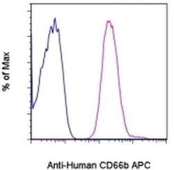
Antigen: CD66b
Classification: Monoclonal
Concentration: 5 μL/Test
Formulation: PBS with 0.2% BSA and 0.09% sodium azide; pH 7.2
Gene Accession No.: P31997
Gene Symbols: CEACAM8
Purification Method: Affinity chromatography
Regulatory Status: RUO
Gene ID (Entrez): 1088
Content And Storage: 4° C, store in dark, DO NOT FREEZE!
Form: Liquid
Applications: Flow Cytometry
Clone: G10F5
Conjugate: APC
Gene: CEACAM8
Gene Alias: carcinoembryonic antigen CGM6; carcinoembryonic antigen gene family member 6; carcinoembryonic antigen related cell adhesion molecule 8; carcinoembryonic antigen-related cell adhesion molecule 8; CD66b; CD67; CD67 antigen; CEACAM8; CGM6; NCA-95; non-specific cross-reacting antigen NCA-95
Host Species: Mouse
Quantity: 100 Tests
Primary or Secondary: Primary
Target Species: Human
Product Type: Antibody
Isotype: IgM κ