50-112-9534
CD279 (PD-1) Monoclonal Antibody (MIH4), eBioscience™, Invitrogen™
Manufacturer: Invitrogen
Select a Size
| Pack Size | SKU | Availability | Price |
|---|---|---|---|
| Each of 1 | 50-112-9534-Each-of-1 | In Stock | ₹ 16,999.00 |
50-112-9534 - Each of 1
In Stock
Quantity
1
Base Price: ₹ 16,999.00
GST (18%): ₹ 3,059.82
Total Price: ₹ 20,058.82
Antigen
CD279 (PD-1)
Classification
Monoclonal
Concentration
0.5 mg/mL
Formulation
PBS with 0.09% sodium azide; pH 7.2
Gene Accession No.
Q15116
Gene Symbols
Pdcd1
Purification Method
Affinity chromatography
Regulatory Status
RUO
Gene ID (Entrez)
5133
Content And Storage
4° C
Form
Liquid
Applications
Flow Cytometry, Immunohistochemistry (Frozen)
Clone
MIH4
Conjugate
Unconjugated
Gene
Pdcd1
Gene Alias
CD279; EGK_05005; hPD1; hPD-1; hPD-l; hSLE1; Ly101; mPD-1; PD1; PD-1; Pdc1; Pdcd1; programmed cell death 1; programmed cell death 1 protein; programmed cell death protein 1; programmed cell death protein 1-like; programmed death 1; Protein PD1; protein PD-1; sCD279; SLEB2; soluble CD279; systemic lupus erythematosus susceptibility 2
Host Species
Mouse
Quantity
100 μg
Primary or Secondary
Primary
Target Species
Human
Product Type
Antibody
Isotype
IgG1 κ
Related Products


Description
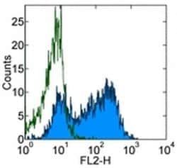
- Description: The MIH4 monoclonal antibody reacts with the human PD-1 (programmed death-1), a 55 kDa member of the immunoglobulin superfamily
- PD-1 contains the immunoreceptor tyrosine-based inhibitory motif (ITIM) and plays a key role in peripheral tolerance and autoimmune disease
- PD-1 is expressed predomitly on activated T and B lymphocytes
- Two novel members of the B7 family have been identified as the PD-1 ligands, PD-L1 (B7-H1) and PD-L2 (B7-DC)
- Evidence reported to date suggests overlapping functions for these two PD-1 ligands and their constitutive expression on some normal tissues and upregulation on activated antigen-presenting cells
- The MIH4 antibody recognizes a different epitope than antibody clones J105
- Applications Reported: This MIH4 antibody has been reported for use in flow cytometric analysis, and immunohistology staining of frozen tissue sections
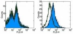
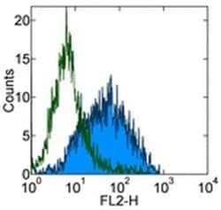
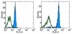
- Applications Tested: This MIH4 antibody has been tested by flow cytometric analysis of PHA-stimulated peripheral blood mononuclear cells
- This can be used at less than or equal to 1 μg per test
- A test is defined as the amount (μg) of antibody that will stain a cell sample in a final volume of 100 μL
- Cell number should be determined empirically but can range from 10^5 to 10^8 cells/test
- It is recommended that the antibody be carefully titrated for optimal performance in the assay of interest
- Purity: Greater than 90%, as determined by SDS-PAGE
- Cell-mediated immune responses are initiated by T lymphocytes that are themselves stimulated by cognate peptides bound to MHC molecules on antig en-presenting cells (APC)
- T-cell activation is generally self-limited as activated T cells express receptors such as PD-1 (also known as PDCD-1) that mediate inhibitory signals from the APC
- PD-1 can bind two different but related ligands, PDL-1 and PDL-2
- Upon binding to either of these ligands, signals generated by PD-1 inhibit the activation of the immune response in the absence of "edanger signals"e such as LPS or other molecules associated with bacteria or other pathogens
- Evidence for this is seen in PD1-null mice who exhibit hyperactivated immune systems and autoimmune diseases
- Despite its predicted molecular weight, PD-1 often migrates at higher molecular weight in SDS-PAGE.
Compare Similar Items
Show Difference
Antigen: CD279 (PD-1)
Classification: Monoclonal
Concentration: 0.5 mg/mL
Formulation: PBS with 0.09% sodium azide; pH 7.2
Gene Accession No.: Q15116
Gene Symbols: Pdcd1
Purification Method: Affinity chromatography
Regulatory Status: RUO
Gene ID (Entrez): 5133
Content And Storage: 4° C
Form: Liquid
Applications: Flow Cytometry, Immunohistochemistry (Frozen)
Clone: MIH4
Conjugate: Unconjugated
Gene: Pdcd1
Gene Alias: CD279; EGK_05005; hPD1; hPD-1; hPD-l; hSLE1; Ly101; mPD-1; PD1; PD-1; Pdc1; Pdcd1; programmed cell death 1; programmed cell death 1 protein; programmed cell death protein 1; programmed cell death protein 1-like; programmed death 1; Protein PD1; protein PD-1; sCD279; SLEB2; soluble CD279; systemic lupus erythematosus susceptibility 2
Host Species: Mouse
Quantity: 100 μg
Primary or Secondary: Primary
Target Species: Human
Product Type: Antibody
Isotype: IgG1 κ
Antigen:
CD279 (PD-1)
Classification:
Monoclonal
Concentration:
0.5 mg/mL
Formulation:
PBS with 0.09% sodium azide; pH 7.2
Gene Accession No.:
Q15116
Gene Symbols:
Pdcd1
Purification Method:
Affinity chromatography
Regulatory Status:
RUO
Gene ID (Entrez):
5133
Content And Storage:
4° C
Form:
Liquid
Applications:
Flow Cytometry, Immunohistochemistry (Frozen)
Clone:
MIH4
Conjugate:
Unconjugated
Gene:
Pdcd1
Gene Alias:
CD279; EGK_05005; hPD1; hPD-1; hPD-l; hSLE1; Ly101; mPD-1; PD1; PD-1; Pdc1; Pdcd1; programmed cell death 1; programmed cell death 1 protein; programmed cell death protein 1; programmed cell death protein 1-like; programmed death 1; Protein PD1; protein PD-1; sCD279; SLEB2; soluble CD279; systemic lupus erythematosus susceptibility 2
Host Species:
Mouse
Quantity:
100 μg
Primary or Secondary:
Primary
Target Species:
Human
Product Type:
Antibody
Isotype:
IgG1 κ
Antigen: CD61 (Integrin beta 3)
Classification: Monoclonal
Concentration: 0.2 mg/mL
Formulation: PBS with 0.09% sodium azide; pH 7.2
Gene Accession No.: O54890
Gene Symbols: ITGB3
Purification Method: Affinity chromatography
Regulatory Status: RUO
Gene ID (Entrez): 16416, 29302
Content And Storage: 4° C, store in dark, DO NOT FREEZE!
Form: Liquid
Applications: Flow Cytometry
Clone: 2C9.G3
Conjugate: PE
Gene: ITGB3
Gene Alias: antigen CD61; BDPLT16; BDPLT2; beta3 integrin; CD61; EGK_08849; glycoprotein GPIIIa; glycoprotein IIIa; GP3A; GPIIIa; GT; INGRB3; integrin beta 3; integrin beta 3 (Cd61); integrin beta-3; integrin beta-3 subunit; integrin subunit beta 3; integrin, beta 3; integrin, beta 3 (platelet glycoprotein IIIa, antigen CD61); ITGB3; platelet glycoprotein IIIa; platelet glycoprotein IIIa integrin subunit beta 3; platelet gpIIIa; platelet membrane glycoprotein IIIa
Host Species: Armenian Hamster
Quantity: 50 μg
Primary or Secondary: Primary
Target Species: Mouse, Rat
Product Type: Antibody
Isotype: IgG
Antigen:
CD61 (Integrin beta 3)
Classification:
Monoclonal
Concentration:
0.2 mg/mL
Formulation:
PBS with 0.09% sodium azide; pH 7.2
Gene Accession No.:
O54890
Gene Symbols:
ITGB3
Purification Method:
Affinity chromatography
Regulatory Status:
RUO
Gene ID (Entrez):
16416, 29302
Content And Storage:
4° C, store in dark, DO NOT FREEZE!
Form:
Liquid
Applications:
Flow Cytometry
Clone:
2C9.G3
Conjugate:
PE
Gene:
ITGB3
Gene Alias:
antigen CD61; BDPLT16; BDPLT2; beta3 integrin; CD61; EGK_08849; glycoprotein GPIIIa; glycoprotein IIIa; GP3A; GPIIIa; GT; INGRB3; integrin beta 3; integrin beta 3 (Cd61); integrin beta-3; integrin beta-3 subunit; integrin subunit beta 3; integrin, beta 3; integrin, beta 3 (platelet glycoprotein IIIa, antigen CD61); ITGB3; platelet glycoprotein IIIa; platelet glycoprotein IIIa integrin subunit beta 3; platelet gpIIIa; platelet membrane glycoprotein IIIa
Host Species:
Armenian Hamster
Quantity:
50 μg
Primary or Secondary:
Primary
Target Species:
Mouse, Rat
Product Type:
Antibody
Isotype:
IgG

Antigen: Rat IgG2a kappa
Classification: __
Concentration: 0.2 mg/mL
Formulation: PBS with 0.09% sodium azide; pH 7.2
Gene Accession No.: __
Gene Symbols: __
Purification Method: Affinity chromatography
Regulatory Status: RUO
Gene ID (Entrez): __
Content And Storage: 4° C, store in dark, DO NOT FREEZE!
Form: Liquid
Applications: Control, Flow Cytometry, Immunocytochemistry, Immunohistochemistry, Immunohistochemistry
Clone: eBR2a
Conjugate: eFluor 660
Gene: __
Gene Alias: IgG2; Immunoglobulin G; Immunoglobulin G2; ImmunoglobulinG; ImmunoglobulinG2
Host Species: Rat
Quantity: 100 μg
Primary or Secondary: Isotype Control (Primary)
Target Species: Not Applicable
Product Type: __
Isotype: IgG2a κ
Antigen:
Rat IgG2a kappa
Classification:
__
Concentration:
0.2 mg/mL
Formulation:
PBS with 0.09% sodium azide; pH 7.2
Gene Accession No.:
__
Gene Symbols:
__
Purification Method:
Affinity chromatography
Regulatory Status:
RUO
Gene ID (Entrez):
__
Content And Storage:
4° C, store in dark, DO NOT FREEZE!
Form:
Liquid
Applications:
Control, Flow Cytometry, Immunocytochemistry, Immunohistochemistry, Immunohistochemistry
Clone:
eBR2a
Conjugate:
eFluor 660
Gene:
__
Gene Alias:
IgG2; Immunoglobulin G; Immunoglobulin G2; ImmunoglobulinG; ImmunoglobulinG2
Host Species:
Rat
Quantity:
100 μg
Primary or Secondary:
Isotype Control (Primary)
Target Species:
Not Applicable
Product Type:
__
Isotype:
IgG2a κ
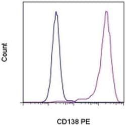
Antigen: CD138 (Syndecan-1)
Classification: Monoclonal
Concentration: 5 μL/Test
Formulation: PBS with 0.2% BSA and 0.09% sodium azide; pH 7.2
Gene Accession No.: P18827
Gene Symbols: Sdc1
Purification Method: Affinity chromatography
Regulatory Status: RUO
Gene ID (Entrez): 6382
Content And Storage: 4° C, store in dark, DO NOT FREEZE!
Form: Liquid
Applications: Flow Cytometry
Clone: DL-101
Conjugate: PE
Gene: Sdc1
Gene Alias: AA408134; AA409076; CD antigen 138; CD138; CD138 antigen; heparan sulfate proteoglycan fibroblast growth factor receptor; HSPG; sCD138; SDC; Sdc1; soluble CD138; Sstn; syn-1; Synd; SYND1; Synd-1; SYNDECA; syndecan; syndecan 1; syndecan proteoglycan 1; Syndecan1; syndecan-1; synstatin
Host Species: Mouse
Quantity: 100 Tests
Primary or Secondary: Primary
Target Species: Human
Product Type: Antibody
Isotype: IgG1 κ
Antigen:
CD138 (Syndecan-1)
Classification:
Monoclonal
Concentration:
5 μL/Test
Formulation:
PBS with 0.2% BSA and 0.09% sodium azide; pH 7.2
Gene Accession No.:
P18827
Gene Symbols:
Sdc1
Purification Method:
Affinity chromatography
Regulatory Status:
RUO
Gene ID (Entrez):
6382
Content And Storage:
4° C, store in dark, DO NOT FREEZE!
Form:
Liquid
Applications:
Flow Cytometry
Clone:
DL-101
Conjugate:
PE
Gene:
Sdc1
Gene Alias:
AA408134; AA409076; CD antigen 138; CD138; CD138 antigen; heparan sulfate proteoglycan fibroblast growth factor receptor; HSPG; sCD138; SDC; Sdc1; soluble CD138; Sstn; syn-1; Synd; SYND1; Synd-1; SYNDECA; syndecan; syndecan 1; syndecan proteoglycan 1; Syndecan1; syndecan-1; synstatin
Host Species:
Mouse
Quantity:
100 Tests
Primary or Secondary:
Primary
Target Species:
Human
Product Type:
Antibody
Isotype:
IgG1 κ