50-153-64
CD279 (PD-1) Monoclonal Antibody (MIH4), APC, eBioscience™, Invitrogen™
Manufacturer: Fischer Scientific
Select a Size
| Pack Size | SKU | Availability | Price |
|---|---|---|---|
| Each of 1 | 50-153-64-Each-of-1 | In Stock | ₹ 17,177.00 |
50-153-64 - Each of 1
In Stock
Quantity
1
Base Price: ₹ 17,177.00
GST (18%): ₹ 3,091.86
Total Price: ₹ 20,268.86
Antigen
CD279 (PD-1)
Classification
Monoclonal
Concentration
5 μL/Test
Formulation
PBS with 0.2% BSA and 0.09% sodium azide; pH 7.2
Gene Accession No.
Q15116
Gene Symbols
Pdcd1
Purification Method
Affinity chromatography
Regulatory Status
RUO
Gene ID (Entrez)
5133
Content And Storage
4° C, store in dark, DO NOT FREEZE!
Form
Liquid
Applications
Flow Cytometry
Clone
MIH4
Conjugate
APC
Gene
Pdcd1
Gene Alias
CD279; EGK_05005; hPD1; hPD-1; hPD-l; hSLE1; Ly101; mPD-1; PD1; PD-1; Pdc1; Pdcd1; programmed cell death 1; programmed cell death 1 protein; programmed cell death protein 1; programmed cell death protein 1-like; programmed death 1; Protein PD1; protein PD-1; sCD279; SLEB2; soluble CD279; systemic lupus erythematosus susceptibility 2
Host Species
Mouse
Quantity
25 Tests
Primary or Secondary
Primary
Target Species
Human
Product Type
Antibody
Isotype
IgG1 κ
Related Products
Description
- Description: The MIH4 monoclonal antibody reacts with the human PD-1 (programmed death-1), a 55 kDa member of the immunoglobulin superfamily
- PD-1 contains the immunoreceptor tyrosine-based inhibitory motif (ITIM) and plays a key role in peripheral tolerance and autoimmune disease
- PD-1 is expressed predominantly on activated T and B lymphocytes
- Two novel members of the B7 family have been identified as the PD-1 ligands, PD-L1 (B7-H1) and PD-L2 (B7-DC)
- Evidence reported to date suggests overlapping functions for these two PD-1 ligands and their constitutive expression on some normal tissues and upregulation on activated antigen-presenting cells
- The MIH4 antibody recognizes a different epitope than antibody clones J105
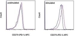
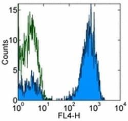
- Applications Reported: This MIH4 antibody has been reported for use in flow cytometric analysis
- Applications Tested: This MIH4 antibody has been pre-titrated and tested by flow cytometric analysis of PHA-stimulated normal human peripheral blood cells
- This can be used at 5 μL (1 μg) per test
- A test is defined as the amount (μg) of antibody that will stain a cell sample in a final volume of 100 μL
- Cell number should be determined empirically but can range from 10^5 to 10^8 cells/test
- Excitation: 633-647 nm; Emission: 660 nm; Laser: Red Laser
- Filtration: 0.2 μm post-manufacturing filtered
- Cell-mediated immune responses are initiated by T lymphocytes that are themselves stimulated by cognate peptides bound to MHC molecules on antig en-presenting cells (APC)
- T-cell activation is generally self-limited as activated T cells express receptors such as PD-1 (also known as PDCD-1) that mediate inhibitory signals from the APC
- PD-1 can bind two different but related ligands, PDL-1 and PDL-2
- Upon binding to either of these ligands, signals generated by PD-1 inhibit the activation of the immune response in the absence of "edanger signals"e such as LPS or other molecules associated with bacteria or other pathogens
- Evidence for this is seen in PD1-null mice who exhibit hyperactivated immune systems and autoimmune diseases
- Despite its predicted molecular weight, PD-1 often migrates at higher molecular weight in SDS-PAGE.
Compare Similar Items
Show Difference
Antigen: CD279 (PD-1)
Classification: Monoclonal
Concentration: 5 μL/Test
Formulation: PBS with 0.2% BSA and 0.09% sodium azide; pH 7.2
Gene Accession No.: Q15116
Gene Symbols: Pdcd1
Purification Method: Affinity chromatography
Regulatory Status: RUO
Gene ID (Entrez): 5133
Content And Storage: 4° C, store in dark, DO NOT FREEZE!
Form: Liquid
Applications: Flow Cytometry
Clone: MIH4
Conjugate: APC
Gene: Pdcd1
Gene Alias: CD279; EGK_05005; hPD1; hPD-1; hPD-l; hSLE1; Ly101; mPD-1; PD1; PD-1; Pdc1; Pdcd1; programmed cell death 1; programmed cell death 1 protein; programmed cell death protein 1; programmed cell death protein 1-like; programmed death 1; Protein PD1; protein PD-1; sCD279; SLEB2; soluble CD279; systemic lupus erythematosus susceptibility 2
Host Species: Mouse
Quantity: 25 Tests
Primary or Secondary: Primary
Target Species: Human
Product Type: Antibody
Isotype: IgG1 κ
Antigen:
CD279 (PD-1)
Classification:
Monoclonal
Concentration:
5 μL/Test
Formulation:
PBS with 0.2% BSA and 0.09% sodium azide; pH 7.2
Gene Accession No.:
Q15116
Gene Symbols:
Pdcd1
Purification Method:
Affinity chromatography
Regulatory Status:
RUO
Gene ID (Entrez):
5133
Content And Storage:
4° C, store in dark, DO NOT FREEZE!
Form:
Liquid
Applications:
Flow Cytometry
Clone:
MIH4
Conjugate:
APC
Gene:
Pdcd1
Gene Alias:
CD279; EGK_05005; hPD1; hPD-1; hPD-l; hSLE1; Ly101; mPD-1; PD1; PD-1; Pdc1; Pdcd1; programmed cell death 1; programmed cell death 1 protein; programmed cell death protein 1; programmed cell death protein 1-like; programmed death 1; Protein PD1; protein PD-1; sCD279; SLEB2; soluble CD279; systemic lupus erythematosus susceptibility 2
Host Species:
Mouse
Quantity:
25 Tests
Primary or Secondary:
Primary
Target Species:
Human
Product Type:
Antibody
Isotype:
IgG1 κ
Antigen: TCR alpha/beta
Classification: Monoclonal
Concentration: 5 μL/Test
Formulation: PBS with 0.2% BSA and 0.09% sodium azide; pH 7.2
Gene Accession No.: P01848, P04435, P0DSE1
Gene Symbols: TRA, TRAC, TRB, TRBV7-9
Purification Method: Affinity chromatography
Regulatory Status: RUO
Gene ID (Entrez): 28589, 28755, 6955, 6957
Content And Storage: 4° C, store in dark, DO NOT FREEZE!
Form: Liquid
Applications: Flow Cytometry
Clone: IP26
Conjugate: APC
Gene: TRAC
Gene Alias: FLJ22602; IMD7; LOC290071; MGC117436; MGC22624; MGC23964; MGC71411; RATTCB; RATTCBC1; RGD1359684; similar to RIKEN cDNA A430107P09 gene; T cell receptor beta locus; T3/TCR complex; TCB; TCBC1; t-cell antigen receptor; T-cell receptor alpha constant; T-cell receptor beta chain; T-cell receptor V alpha; tcr alpha; TCR alpha/ beta; TCR beta; TCRA; Tcrb; TRA; Tra29; TRAC; TRB; TRB@; TRCA
Host Species: Mouse
Quantity: 100 Tests
Primary or Secondary: Primary
Target Species: Human
Product Type: Antibody
Isotype: IgG1 κ
Antigen:
TCR alpha/beta
Classification:
Monoclonal
Concentration:
5 μL/Test
Formulation:
PBS with 0.2% BSA and 0.09% sodium azide; pH 7.2
Gene Accession No.:
P01848, P04435, P0DSE1
Gene Symbols:
TRA, TRAC, TRB, TRBV7-9
Purification Method:
Affinity chromatography
Regulatory Status:
RUO
Gene ID (Entrez):
28589, 28755, 6955, 6957
Content And Storage:
4° C, store in dark, DO NOT FREEZE!
Form:
Liquid
Applications:
Flow Cytometry
Clone:
IP26
Conjugate:
APC
Gene:
TRAC
Gene Alias:
FLJ22602; IMD7; LOC290071; MGC117436; MGC22624; MGC23964; MGC71411; RATTCB; RATTCBC1; RGD1359684; similar to RIKEN cDNA A430107P09 gene; T cell receptor beta locus; T3/TCR complex; TCB; TCBC1; t-cell antigen receptor; T-cell receptor alpha constant; T-cell receptor beta chain; T-cell receptor V alpha; tcr alpha; TCR alpha/ beta; TCR beta; TCRA; Tcrb; TRA; Tra29; TRAC; TRB; TRB@; TRCA
Host Species:
Mouse
Quantity:
100 Tests
Primary or Secondary:
Primary
Target Species:
Human
Product Type:
Antibody
Isotype:
IgG1 κ

Antigen: __
Classification: __
Concentration: __
Formulation: __
Gene Accession No.: __
Gene Symbols: __
Purification Method: __
Regulatory Status: __
Gene ID (Entrez): __
Content And Storage: __
Form: __
Applications: __
Clone: __
Conjugate: __
Gene: __
Gene Alias: __
Host Species: __
Quantity: __
Primary or Secondary: __
Target Species: __
Product Type: __
Isotype: __
Antigen:
__
Classification:
__
Concentration:
__
Formulation:
__
Gene Accession No.:
__
Gene Symbols:
__
Purification Method:
__
Regulatory Status:
__
Gene ID (Entrez):
__
Content And Storage:
__
Form:
__
Applications:
__
Clone:
__
Conjugate:
__
Gene:
__
Gene Alias:
__
Host Species:
__
Quantity:
__
Primary or Secondary:
__
Target Species:
__
Product Type:
__
Isotype:
__
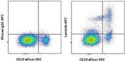
Antigen: Lambda light chain
Classification: Monoclonal
Concentration: 5 μL/Test
Formulation: PBS with 0.2% BSA and 0.09% sodium azide; pH 7.2
Gene Accession No.: P01701, P0CG04
Gene Symbols: IGL, IGLC1, IGLV, IGLV1-51
Purification Method: Affinity chromatography
Regulatory Status: RUO
Gene ID (Entrez): 28820, 3535, 3537, 3546
Content And Storage: 4° C, store in dark, DO NOT FREEZE!
Form: Liquid
Applications: Flow Cytometry
Clone: 1-155-2
Conjugate: APC
Gene: IGLC1
Gene Alias: Bence Jones Protein; BJP; Clambda1; Ig lambda chain V-I region BL2; IgL; Igl@; IGLC; IGLC 1/2/3; IGLC1; Igl-C1; IGLL5; IGLV; IGLV@; immmunoglobulin lambda light chain variable region; Immunoglobulin L; immunoglobulin lambda chain complex; immunoglobulin lambda constant 1; immunoglobulin lambda constant 7; immunoglobulin lambda light chain; immunoglobulin lambda light chain constant region; immunoglobulin lambda light chain V-J region; immunoglobulin lambda variable cluster; Lambda LC; lambda-immunoglobulin; LOC100060608; Mcg Marker; Paraprotein
Host Species: Mouse
Quantity: 100 Tests
Primary or Secondary: Primary
Target Species: Human
Product Type: Antibody
Isotype: IgG1 κ
Antigen:
Lambda light chain
Classification:
Monoclonal
Concentration:
5 μL/Test
Formulation:
PBS with 0.2% BSA and 0.09% sodium azide; pH 7.2
Gene Accession No.:
P01701, P0CG04
Gene Symbols:
IGL, IGLC1, IGLV, IGLV1-51
Purification Method:
Affinity chromatography
Regulatory Status:
RUO
Gene ID (Entrez):
28820, 3535, 3537, 3546
Content And Storage:
4° C, store in dark, DO NOT FREEZE!
Form:
Liquid
Applications:
Flow Cytometry
Clone:
1-155-2
Conjugate:
APC
Gene:
IGLC1
Gene Alias:
Bence Jones Protein; BJP; Clambda1; Ig lambda chain V-I region BL2; IgL; Igl@; IGLC; IGLC 1/2/3; IGLC1; Igl-C1; IGLL5; IGLV; IGLV@; immmunoglobulin lambda light chain variable region; Immunoglobulin L; immunoglobulin lambda chain complex; immunoglobulin lambda constant 1; immunoglobulin lambda constant 7; immunoglobulin lambda light chain; immunoglobulin lambda light chain constant region; immunoglobulin lambda light chain V-J region; immunoglobulin lambda variable cluster; Lambda LC; lambda-immunoglobulin; LOC100060608; Mcg Marker; Paraprotein
Host Species:
Mouse
Quantity:
100 Tests
Primary or Secondary:
Primary
Target Species:
Human
Product Type:
Antibody
Isotype:
IgG1 κ