CD3 epsilon Antibody (B-B12), Novus Biologicals™
Mouse Monoclonal Antibody
Manufacturer: Fischer Scientific
The price for this product is unavailable. Please request a quote
Antigen
CD3 epsilon
Concentration
0.2 mg/mL
Applications
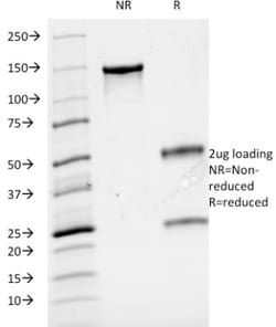
Flow Cytometry, SDS-Page, Immunofluorescence
Conjugate
Unconjugated
Host Species
Mouse
Research Discipline
Adaptive Immunity, Apoptosis, Cytokine Research, Diabetes Research, Immunology, Innate Immunity, Mesenchymal Stem Cell Markers, Signal Transduction, Stem Cell Lines, Stem Cell Markers
Formulation
10mM PBS and 0.05% BSA with 0.05% Sodium Azide
Gene ID (Entrez)
916
Immunogen
Human T cell leukemia cells
Primary or Secondary
Primary
Content And Storage
Store at 4C.
Molecular Weight of Antigen
20 kDa
Clone
B-B12
Dilution
Flow Cytometry 0.5 - 1 ug/million cells in 0.1 ml, SDS-Page, Immunofluorescence 1 - 2 ug/ml
Classification
Monoclonal
Form
Purified
Regulatory Status
RUO
Target Species
Human
Gene Alias
CD3e antigen, CD3e antigen, epsilon polypeptide (TiT3 complex), CD3e molecule, epsilon (CD3-TCR complex), CD3-epsilon, FLJ18683, T3E, T-cell antigen receptor complex, epsilon subunit of T3, T-cell surface antigen T3/Leu-4 epsilon chain, T-cell surface glycoprotein CD3 epsilon chain, TCRE
Gene Symbols
CD3E
Isotype
IgG1 κ
Purification Method
Protein A purified
Test Specificity
Reacts with five invariable CD3 chains (designated as and ) with molecular weight ranging from 16-28kDa. CD3 is expressed, typically at high levels, on peripheral T cells and majority of T cell neoplasms. Thymocytes express CD3 at different level on the cell surface in the course of differentiation and, in cortical thymus, CD3 is predominantly Intracytoplasmic. The CD3 complex is closely associated at the lymphocyte cell surface with T cell antigen receptor (TCR) and is involved in transducing antigen-recognition signals into cytoplasm of T cells and in regulating the cell surface expression of the TCR complex.
Description
- Ensure accurate, reproducible results in Flow Cytometry, Immunofluorescence CD3 epsilon Monoclonal specifically detects CD3 epsilon in Human samples
- It is validated for Flow Cytometry, Immunocytochemistry/Immunofluorescence.
Compare Similar Items
Show Difference
Antigen: CD3 epsilon
Concentration: 0.2 mg/mL
Applications: Flow Cytometry, SDS-Page, Immunofluorescence
Conjugate: Unconjugated
Host Species: Mouse
Research Discipline: Adaptive Immunity, Apoptosis, Cytokine Research, Diabetes Research, Immunology, Innate Immunity, Mesenchymal Stem Cell Markers, Signal Transduction, Stem Cell Lines, Stem Cell Markers
Formulation: 10mM PBS and 0.05% BSA with 0.05% Sodium Azide
Gene ID (Entrez): 916
Immunogen: Human T cell leukemia cells
Primary or Secondary: Primary
Content And Storage: Store at 4C.
Molecular Weight of Antigen: 20 kDa
Clone: B-B12
Dilution: Flow Cytometry 0.5 - 1 ug/million cells in 0.1 ml, SDS-Page, Immunofluorescence 1 - 2 ug/ml
Classification: Monoclonal
Form: Purified
Regulatory Status: RUO
Target Species: Human
Gene Alias: CD3e antigen, CD3e antigen, epsilon polypeptide (TiT3 complex), CD3e molecule, epsilon (CD3-TCR complex), CD3-epsilon, FLJ18683, T3E, T-cell antigen receptor complex, epsilon subunit of T3, T-cell surface antigen T3/Leu-4 epsilon chain, T-cell surface glycoprotein CD3 epsilon chain, TCRE
Gene Symbols: CD3E
Isotype: IgG1 κ
Purification Method: Protein A purified
Test Specificity: Reacts with five invariable CD3 chains (designated as and ) with molecular weight ranging from 16-28kDa. CD3 is expressed, typically at high levels, on peripheral T cells and majority of T cell neoplasms. Thymocytes express CD3 at different level on the cell surface in the course of differentiation and, in cortical thymus, CD3 is predominantly Intracytoplasmic. The CD3 complex is closely associated at the lymphocyte cell surface with T cell antigen receptor (TCR) and is involved in transducing antigen-recognition signals into cytoplasm of T cells and in regulating the cell surface expression of the TCR complex.

Antigen: CD4
Concentration: 0.2 mg/mL
Applications: Flow Cytometry, SDS-Page, Immunofluorescence
Conjugate: Unconjugated
Host Species: Mouse
Research Discipline: Adaptive Immunity, Cell Biology, Cellular Markers, Cytokine Research, Immunology, Innate Immunity, Regulatory Immunology, Stem Cell Markers
Formulation: 10mM PBS and 0.05% BSA with 0.05% Sodium Azide
Gene ID (Entrez): 920
Immunogen: Recombinant human CD4 protein
Primary or Secondary: Primary
Content And Storage: Store at 4C.
Molecular Weight of Antigen: 55 kDa
Clone: C4/206
Dilution: Flow Cytometry 0.5 - 1 ug/million cells in 0.1 ml, SDS-Page, Immunofluorescence 0.5 - 1.0 ug/ml
Classification: Monoclonal
Form: Purified
Regulatory Status: RUO
Target Species: Human
Gene Alias: CD4 antigen, CD4 antigen (p55), CD4 molecule, CD4 receptor, CD4mut, T-cell surface antigen T4/Leu-3, T-cell surface glycoprotein CD4
Gene Symbols: __
Isotype: IgG1 κ
Purification Method: Protein A or G purified
Test Specificity: Recognizes a protein of 55kDa, identified as CD4. CD4 is a membrane glycoprotein of T lymphocytes that interacts with major histocompatibility complex class II antigens and is also a receptor for the human immunodeficiency virus. This protein is expressed not only in T lymphocytes, but also in B cells, macrophages, and granulocytes. It is also expressed in specific regions of the brain. The protein functions to initiate or augment the early phase of T-cell activation, and may function as an important mediator of indirect neuronal damage in infectious and immune-mediated diseases of the central nervous system. Multiple alternatively spliced transcript variants encoding different isoforms have been identified