CD4 Antibody (EDU-2), Novus Biologicals™
Mouse Monoclonal Antibody
Manufacturer: Fischer Scientific
The price for this product is unavailable. Please request a quote
Antigen
CD4
Concentration
0.2 mg/mL
Applications
Flow Cytometry, Immunofluorescence
Conjugate
Unconjugated
Host Species
Mouse
Research Discipline
Adaptive Immunity, Cell Biology, Cellular Markers, Cytokine Research, Immunology, Innate Immunity, Regulatory Immunology, Stem Cell Markers
Formulation
10mM PBS and 0.05% BSA with 0.05% Sodium Azide
Gene ID (Entrez)
920
Isotype
IgG2a κ
Purification Method
Protein A or G purified
Test Specificity
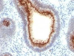
Recognizes a protein of 55kDa, identified as CD4. It is a membrane glycoprotein of T lymphocytes that interacts with major histocompatibility complex class II antigens and is also a receptor for the human immunodeficiency virus. This protein is expressed not only in T lymphocytes, but also in B cells, macrophages, and granulocytes. It is also expressed in specific regions of the brain. The protein functions to initiate or augment the early phase of T-cell activation, and may function as an important mediator of indirect neuronal damage in infectious and immune-mediated diseases of the central nervous system. Multiple alternatively spliced transcript variants encoding different isoforms have been identified. This MAb was characterized as human CD4 antibody at II and IV International Workshop on Human Leukocyte Differentiation Antigens.
Clone
EDU-2
Dilution
Flow Cytometry 0.5 - 1 ug/million cells in 0.1 ml, Immunofluorescence 0.5 - 1.0 ug/ml
Classification
Monoclonal
Form
Purified
Regulatory Status
RUO
Target Species
Human, Monkey, Orangutan
Gene Alias
CD4 antigen, CD4 antigen (p55), CD4 molecule, CD4 receptor, CD4mut, T-cell surface antigen T4/Leu-3, T-cell surface glycoprotein CD4
Immunogen
Stimulated human leukocytes
Primary or Secondary
Primary
Content And Storage
Store at 4C.
Molecular Weight of Antigen
55 kDa
Description
- Ensure accurate, reproducible results in Flow Cytometry, Immunofluorescence CD4 Monoclonal specifically detects CD4 in Human, Monkey, Chimpanzee, Orangutan samples
- It is validated for Flow Cytometry, Immunocytochemistry/Immunofluorescence, Functional, Immunofluorescence.
Compare Similar Items
Show Difference

Antigen: CD4
Concentration: 0.2 mg/mL
Applications: Flow Cytometry, Immunofluorescence
Conjugate: Unconjugated
Host Species: Mouse
Research Discipline: Adaptive Immunity, Cell Biology, Cellular Markers, Cytokine Research, Immunology, Innate Immunity, Regulatory Immunology, Stem Cell Markers
Formulation: 10mM PBS and 0.05% BSA with 0.05% Sodium Azide
Gene ID (Entrez): 920
Isotype: IgG2a κ
Purification Method: Protein A or G purified
Test Specificity: Recognizes a protein of 55kDa, identified as CD4. It is a membrane glycoprotein of T lymphocytes that interacts with major histocompatibility complex class II antigens and is also a receptor for the human immunodeficiency virus. This protein is expressed not only in T lymphocytes, but also in B cells, macrophages, and granulocytes. It is also expressed in specific regions of the brain. The protein functions to initiate or augment the early phase of T-cell activation, and may function as an important mediator of indirect neuronal damage in infectious and immune-mediated diseases of the central nervous system. Multiple alternatively spliced transcript variants encoding different isoforms have been identified. This MAb was characterized as human CD4 antibody at II and IV International Workshop on Human Leukocyte Differentiation Antigens.
Clone: EDU-2
Dilution: Flow Cytometry 0.5 - 1 ug/million cells in 0.1 ml, Immunofluorescence 0.5 - 1.0 ug/ml
Classification: Monoclonal
Form: Purified
Regulatory Status: RUO
Target Species: Human, Monkey, Orangutan
Gene Alias: CD4 antigen, CD4 antigen (p55), CD4 molecule, CD4 receptor, CD4mut, T-cell surface antigen T4/Leu-3, T-cell surface glycoprotein CD4
Immunogen: Stimulated human leukocytes
Primary or Secondary: Primary
Content And Storage: Store at 4C.
Molecular Weight of Antigen: 55 kDa
Antigen: CDC2/CDK1
Concentration: 0.2 mg/mL
Applications: Flow Cytometry, Immunohistochemistry (Paraffin), Immunofluorescence
Conjugate: Unconjugated
Host Species: Mouse
Research Discipline: Breast Cancer, Cancer, Cell Cycle and Replication, Core ESC Like Genes, Mitotic Regulators, Stem Cell Markers
Formulation: 10mM PBS and 0.05% BSA with 0.05% Sodium Azide
Gene ID (Entrez): 983
Isotype: IgG2a κ
Purification Method: Protein A or G purified
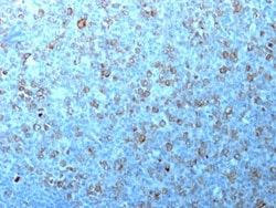
Test Specificity: Recognizes a 34kDa protein, which is identified as cyclin dependent kinase 1 (cdk1) or p34cdc2 protein kinase. cdk1/ p34cdc2 plays a crucial role during cell division and is most active during mitosis. It is predominantly localized in the nucleus. It is a serine/threonine kinase, which is activated by cyclin, presumably by de-phosphorylation of tyrosine residues. Activated cdk1/ p34cdc2 performs specific functions during mitosis, including nuclear envelope breakdown and chromosome condensation.
Clone: CDK1/873
Dilution: Flow Cytometry 0.5 - 1 ug/million cells in 0.1 ml, Immunohistochemistry-Paraffin 2 - 4 ug/ml, Immunofluorescence 1 - 2 ug/ml
Classification: Monoclonal
Form: Purified
Regulatory Status: RUO
Target Species: Human, Monkey, Mink, Primate, S. pombe (Negative), Drosophila (Negative), Mouse (Negative), Rat (Negative)
Gene Alias: CDC28A, CDC2MGC111195, cell cycle controller CDC2, Cell division control protein 2 homolog, Cell division protein kinase 1, cyclin-dependent kinase 1, DKFZp686L20222, EC 2.7.11.22, EC 2.7.11.23, G1 to S and G2 to M, p34 protein kinase, P34CDC2
Immunogen: Recombinant human CDK1 protein
Primary or Secondary: Primary
Content And Storage: Store at 4C.
Molecular Weight of Antigen: 34 kDa
Antigen: Cdc20
Concentration: 0.2mg/mL
Applications: Flow Cytometry, Immunohistochemistry (Paraffin), Immunofluorescence
Conjugate: Unconjugated
Host Species: Mouse
Research Discipline: Cell Cycle and Replication, Core ESC Like Genes, Stem Cell Markers
Formulation: 10mM PBS and 0.05% BSA with 0.05% Sodium Azide
Gene ID (Entrez): 991
Isotype: IgG1 κ
Purification Method: Protein A or G purified
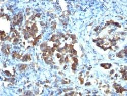
Test Specificity: Cyclins, regulatory subunits, which associate with kinases, control many of the important steps in cell cycle progression. The Cdc2 protein kinase (p34Cdc2) exhibits protein kinase activity in vitro and exists in a complex with both cyclin B and a protein homologous to p13SUC1. Cdc2 kinase is the active subunit of the M phase promoting factor (MPF) and the M phase-specific Histone H1 kinase. The p34Cdc2/cyclin B complex is required for the G2 to M transition. An additional cell cycle-dependent protein kinase, termed p55cdc, exhibits a high degree of homology with the S. cerevisiae proteins Cdc20 and Cdc4. The p55cdc transcript is readily detectable in a variety of cultured cell lines in growth phase, but disappears when cell growth is chemically arrested.
Clone: CDC20/1102
Dilution: Flow Cytometry 0.5 - 1 ug/million cells in 0.1 ml, Immunohistochemistry-Paraffin 2 - 4 ug/ml, Immunofluorescence 0.5 - 1.0 ug/ml
Classification: Monoclonal
Form: Purified
Regulatory Status: RUO
Target Species: Human
Gene Alias: CDC20 cell division cycle 20 homolog, CDC20 cell division cycle 20 homolog (S. cerevisiae), cell division cycle 20 homolog (S. cerevisiae), cell division cycle protein 20 homolog, MGC102824, p55CDCS. cerevisiae, homolog)
Immunogen: Recombinant human Cdc20 protein
Primary or Secondary: Primary
Content And Storage: Store at 4C.
Molecular Weight of Antigen: 55 kDa