CDC2/CDK1 Antibody (CDK1/873), Novus Biologicals™
Mouse Monoclonal Antibody
Manufacturer: Fischer Scientific
The price for this product is unavailable. Please request a quote
Antigen
CDC2/CDK1
Concentration
0.2 mg/mL
Applications
Flow Cytometry, Immunohistochemistry (Paraffin), Immunofluorescence
Conjugate
Unconjugated
Host Species
Mouse
Research Discipline
Breast Cancer, Cancer, Cell Cycle and Replication, Core ESC Like Genes, Mitotic Regulators, Stem Cell Markers
Formulation
10mM PBS and 0.05% BSA with 0.05% Sodium Azide
Gene ID (Entrez)
983
Immunogen
Recombinant human CDK1 protein
Primary or Secondary
Primary
Content And Storage
Store at 4C.
Molecular Weight of Antigen
34 kDa
Clone
CDK1/873
Dilution
Flow Cytometry 0.5 - 1 ug/million cells in 0.1 ml, Immunohistochemistry-Paraffin 2 - 4 ug/ml, Immunofluorescence 1 - 2 ug/ml
Classification
Monoclonal
Form
Purified
Regulatory Status
RUO
Target Species
Human, Monkey, Mink, Primate, S. pombe (Negative), Drosophila (Negative), Mouse (Negative), Rat (Negative)
Gene Alias
CDC28A, CDC2MGC111195, cell cycle controller CDC2, Cell division control protein 2 homolog, Cell division protein kinase 1, cyclin-dependent kinase 1, DKFZp686L20222, EC 2.7.11.22, EC 2.7.11.23, G1 to S and G2 to M, p34 protein kinase, P34CDC2
Gene Symbols
CDK1
Isotype
IgG2a κ
Purification Method
Protein A or G purified
Test Specificity
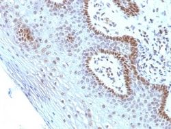
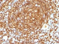
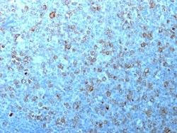
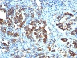
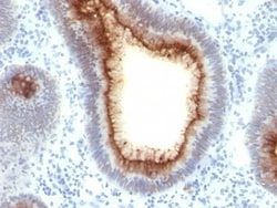
Recognizes a 34kDa protein, which is identified as cyclin dependent kinase 1 (cdk1) or p34cdc2 protein kinase. cdk1/ p34cdc2 plays a crucial role during cell division and is most active during mitosis. It is predominantly localized in the nucleus. It is a serine/threonine kinase, which is activated by cyclin, presumably by de-phosphorylation of tyrosine residues. Activated cdk1/ p34cdc2 performs specific functions during mitosis, including nuclear envelope breakdown and chromosome condensation.
Related Products


Description
- Ensure accurate, reproducible results in Flow Cytometry, Immunohistochemistry (Paraffin), Immunofluorescence CDC2/CDK1 Monoclonal specifically detects CDC2/CDK1 in Human, Bovine, Mink, Monkey, S
- pombe (Negative), Drosophila (Negative), Mouse (Negative), Rat (Negative), Xenopus (Negative), Yeast (Negative) samples
- It is validated for Western Blot, Immunohistochemistry, Immunohistochemistry-Paraffin.
Compare Similar Items
Show Difference
Antigen: CDC2/CDK1
Concentration: 0.2 mg/mL
Applications: Flow Cytometry, Immunohistochemistry (Paraffin), Immunofluorescence
Conjugate: Unconjugated
Host Species: Mouse
Research Discipline: Breast Cancer, Cancer, Cell Cycle and Replication, Core ESC Like Genes, Mitotic Regulators, Stem Cell Markers
Formulation: 10mM PBS and 0.05% BSA with 0.05% Sodium Azide
Gene ID (Entrez): 983
Immunogen: Recombinant human CDK1 protein
Primary or Secondary: Primary
Content And Storage: Store at 4C.
Molecular Weight of Antigen: 34 kDa
Clone: CDK1/873
Dilution: Flow Cytometry 0.5 - 1 ug/million cells in 0.1 ml, Immunohistochemistry-Paraffin 2 - 4 ug/ml, Immunofluorescence 1 - 2 ug/ml
Classification: Monoclonal
Form: Purified
Regulatory Status: RUO
Target Species: Human, Monkey, Mink, Primate, S. pombe (Negative), Drosophila (Negative), Mouse (Negative), Rat (Negative)
Gene Alias: CDC28A, CDC2MGC111195, cell cycle controller CDC2, Cell division control protein 2 homolog, Cell division protein kinase 1, cyclin-dependent kinase 1, DKFZp686L20222, EC 2.7.11.22, EC 2.7.11.23, G1 to S and G2 to M, p34 protein kinase, P34CDC2
Gene Symbols: CDK1
Isotype: IgG2a κ
Purification Method: Protein A or G purified
Test Specificity: Recognizes a 34kDa protein, which is identified as cyclin dependent kinase 1 (cdk1) or p34cdc2 protein kinase. cdk1/ p34cdc2 plays a crucial role during cell division and is most active during mitosis. It is predominantly localized in the nucleus. It is a serine/threonine kinase, which is activated by cyclin, presumably by de-phosphorylation of tyrosine residues. Activated cdk1/ p34cdc2 performs specific functions during mitosis, including nuclear envelope breakdown and chromosome condensation.
Antigen: Cdc20
Concentration: 0.2mg/mL
Applications: Flow Cytometry, Immunohistochemistry (Paraffin), Immunofluorescence
Conjugate: Unconjugated
Host Species: Mouse
Research Discipline: Cell Cycle and Replication, Core ESC Like Genes, Stem Cell Markers
Formulation: 10mM PBS and 0.05% BSA with 0.05% Sodium Azide
Gene ID (Entrez): 991
Immunogen: Recombinant human Cdc20 protein
Primary or Secondary: Primary
Content And Storage: Store at 4C.
Molecular Weight of Antigen: 55 kDa
Clone: CDC20/1102
Dilution: Flow Cytometry 0.5 - 1 ug/million cells in 0.1 ml, Immunohistochemistry-Paraffin 2 - 4 ug/ml, Immunofluorescence 0.5 - 1.0 ug/ml
Classification: Monoclonal
Form: Purified
Regulatory Status: RUO
Target Species: Human
Gene Alias: CDC20 cell division cycle 20 homolog, CDC20 cell division cycle 20 homolog (S. cerevisiae), cell division cycle 20 homolog (S. cerevisiae), cell division cycle protein 20 homolog, MGC102824, p55CDCS. cerevisiae, homolog)
Gene Symbols: CDC20
Isotype: IgG1 κ
Purification Method: Protein A or G purified
Test Specificity: Cyclins, regulatory subunits, which associate with kinases, control many of the important steps in cell cycle progression. The Cdc2 protein kinase (p34Cdc2) exhibits protein kinase activity in vitro and exists in a complex with both cyclin B and a protein homologous to p13SUC1. Cdc2 kinase is the active subunit of the M phase promoting factor (MPF) and the M phase-specific Histone H1 kinase. The p34Cdc2/cyclin B complex is required for the G2 to M transition. An additional cell cycle-dependent protein kinase, termed p55cdc, exhibits a high degree of homology with the S. cerevisiae proteins Cdc20 and Cdc4. The p55cdc transcript is readily detectable in a variety of cultured cell lines in growth phase, but disappears when cell growth is chemically arrested.
Antigen: CEACAM5/CD66e
Concentration: 0.2mg/mL
Applications: Flow Cytometry, Immunohistochemistry (Paraffin), SDS-Page, Immunofluorescence
Conjugate: Unconjugated
Host Species: Mouse
Research Discipline: Cancer, Cellular Markers, Immunology
Formulation: 10mM PBS and 0.05% BSA with 0.05% Sodium Azide
Gene ID (Entrez): 1048
Immunogen: Recombinant full-length human CEA protein
Primary or Secondary: Primary
Content And Storage: Store at 4C.
Molecular Weight of Antigen: __
Clone: C66/1009
Dilution: Flow Cytometry 0.5 - 1 ug/million cells in 0.1 ml, Immunohistochemistry-Paraffin 0.5 - 1.0 ug/ml, SDS-Page, Immunofluorescence 1 - 2 ug/ml
Classification: Monoclonal
Form: Purified
Regulatory Status: RUO
Target Species: Human
Gene Alias: Carcinoembryonic antigen, carcinoembryonic antigen-related cell adhesion molecule 5, CD66e antigen, CEACD66e, DKFZp781M2392, Meconium antigen 100
Gene Symbols: CEACAM5
Isotype: IgG2a κ
Purification Method: Protein A or G purified
Test Specificity: This antibody recognizes proteins of 80-200kDa, identified as different members of CEA family. CEA is synthesized during development in the fetal gut and is re-expressed in increased amounts in intestinal carcinomas and several other tumors. This MAb does not react with nonspecific cross-reacting antigen (NCA) and with human polymorphonuclear leucocytes. It shows no reaction with a variety of normal tissues and is suitable for staining of formalin/paraffin tissues. CEA is not found in benign glands, stroma, or malignant prostatic cells. Antibody to CEA is useful in detecting early foci of gastric carcinoma and in distinguishing pulmonary adenocarcinomas (60-70% are CEA+) from pleural mesotheliomas (rarely or weakly CEA+). Anti-CEA positivity is seen in adenocarcinomas from the lung, colon, stomach, esophagus, pancreas, gallbadder, urachus, salivary gland, ovary, and endocervix.