CD7 Antibody (T3-3A1), Novus Biologicals™
Manufacturer: Fischer Scientific
Select a Size
| Pack Size | SKU | Availability | Price |
|---|---|---|---|
| Each of 1 | NBP24480300-Each-of-1 | In Stock | ₹ 24,920.00 |
NBP24480300 - Each of 1
In Stock
Quantity
1
Base Price: ₹ 24,920.00
GST (18%): ₹ 4,485.60
Total Price: ₹ 29,405.60
Antigen
CD7
Classification
Monoclonal
Concentration
0.2 mg/mL
Dilution
Flow Cytometry 0.5 - 1 ug/million cells in 0.1 ml, Immunofluorescence 0.5 - 1.0 ug/ml
Gene Alias
CD7 antigen, CD7 antigen (p41), CD7 molecule, GP40T-cell surface antigen Leu-9, LEU-9, T-cell antigen CD7, T-cell leukemia antigen, Tp40, TP41p41 protein
Host Species
Mouse
Molecular Weight of Antigen
40 kDa
Quantity
0.02 mg
Research Discipline
Cytokine Research, Signal Transduction
Gene ID (Entrez)
924
Target Species
Human
Form
Purified
Applications
Flow Cytometry, Immunofluorescence
Clone
T3-3A1
Conjugate
Unconjugated
Formulation
10mM PBS and 0.05% BSA with 0.05% Sodium Azide
Gene Symbols
CD7
Immunogen
Human T cells
Purification Method
Protein A or G purified
Regulatory Status
RUO
Primary or Secondary
Primary
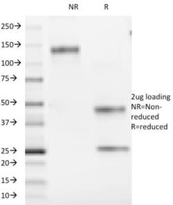
Test Specificity
Recognizes a protein of 40kDa, identified as CD7, a member of the immunoglobulin gene superfamily. Its N-terminal amino acids 1-107 are highly homologous to Ig kappa-L chains whereas the carboxyl-terminal region of the extracellular domain is proline-rich and has been postulated to form a stalk from which the Ig domain projects. CD7 is expressed on the majority of immature and mature T-lymphocytes, and T cell leukemia. It is also found on natural killer cells, a small subpopulation of normal B cells and on malignant B cells. Cross-linking surface CD7 positively modulates T cell and NK cell activity as measured by calcium fluxes, expression of adhesion molecules, cytokine secretion and proliferation. CD7 associates directly with phosphoinositol 3'-kinase. CD7 ligation induces production of D-3 phosphoinositides and tyrosine phosphorylation.
Content And Storage
Store at 4C.
Isotype
IgG1 κ
Related Products


Description
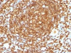
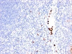
- Ensure accurate, reproducible results in Flow Cytometry, Immunofluorescence CD7 Monoclonal specifically detects CD7 in Human samples
- It is validated for Flow Cytometry, Immunocytochemistry/Immunofluorescence, Immunofluorescence.
Compare Similar Items
Show Difference
Antigen: CD7
Classification: Monoclonal
Concentration: 0.2 mg/mL
Dilution: Flow Cytometry 0.5 - 1 ug/million cells in 0.1 ml, Immunofluorescence 0.5 - 1.0 ug/ml
Gene Alias: CD7 antigen, CD7 antigen (p41), CD7 molecule, GP40T-cell surface antigen Leu-9, LEU-9, T-cell antigen CD7, T-cell leukemia antigen, Tp40, TP41p41 protein
Host Species: Mouse
Molecular Weight of Antigen: 40 kDa
Quantity: 0.02 mg
Research Discipline: Cytokine Research, Signal Transduction
Gene ID (Entrez): 924
Target Species: Human
Form: Purified
Applications: Flow Cytometry, Immunofluorescence
Clone: T3-3A1
Conjugate: Unconjugated
Formulation: 10mM PBS and 0.05% BSA with 0.05% Sodium Azide
Gene Symbols: CD7
Immunogen: Human T cells
Purification Method: Protein A or G purified
Regulatory Status: RUO
Primary or Secondary: Primary
Test Specificity: Recognizes a protein of 40kDa, identified as CD7, a member of the immunoglobulin gene superfamily. Its N-terminal amino acids 1-107 are highly homologous to Ig kappa-L chains whereas the carboxyl-terminal region of the extracellular domain is proline-rich and has been postulated to form a stalk from which the Ig domain projects. CD7 is expressed on the majority of immature and mature T-lymphocytes, and T cell leukemia. It is also found on natural killer cells, a small subpopulation of normal B cells and on malignant B cells. Cross-linking surface CD7 positively modulates T cell and NK cell activity as measured by calcium fluxes, expression of adhesion molecules, cytokine secretion and proliferation. CD7 associates directly with phosphoinositol 3'-kinase. CD7 ligation induces production of D-3 phosphoinositides and tyrosine phosphorylation.
Content And Storage: Store at 4C.
Isotype: IgG1 κ
Antigen: CD7
Classification: Monoclonal
Concentration: 0.2 mg/mL
Dilution: Flow Cytometry 0.5 - 1 ug/million cells in 0.1 ml, Immunofluorescence 0.5 - 1.0 ug/ml
Gene Alias: CD7 antigen, CD7 antigen (p41), CD7 molecule, GP40T-cell surface antigen Leu-9, LEU-9, T-cell antigen CD7, T-cell leukemia antigen, Tp40, TP41p41 protein
Host Species: Mouse
Molecular Weight of Antigen: 40 kDa
Quantity: 0.1 mg
Research Discipline: Cytokine Research, Signal Transduction
Gene ID (Entrez): 924
Target Species: Human
Form: Purified
Applications: Flow Cytometry, Immunofluorescence
Clone: T3-3A1
Conjugate: Unconjugated
Formulation: 10mM PBS and 0.05% BSA with 0.05% Sodium Azide
Gene Symbols: CD7
Immunogen: Human T cells
Purification Method: Protein A or G purified
Regulatory Status: RUO
Primary or Secondary: Primary
Test Specificity: Recognizes a protein of 40kDa, identified as CD7, a member of the immunoglobulin gene superfamily. Its N-terminal amino acids 1-107 are highly homologous to Ig kappa-L chains whereas the carboxyl-terminal region of the extracellular domain is proline-rich and has been postulated to form a stalk from which the Ig domain projects. CD7 is expressed on the majority of immature and mature T-lymphocytes, and T cell leukemia. It is also found on natural killer cells, a small subpopulation of normal B cells and on malignant B cells. Cross-linking surface CD7 positively modulates T cell and NK cell activity as measured by calcium fluxes, expression of adhesion molecules, cytokine secretion and proliferation. CD7 associates directly with phosphoinositol 3'-kinase. CD7 ligation induces production of D-3 phosphoinositides and tyrosine phosphorylation.
Content And Storage: Store at 4C.
Isotype: IgG1 κ
Antigen: CD48/SLAMF2
Classification: Monoclonal
Concentration: 0.2 mg/mL
Dilution: Flow Cytometry 0.5 - 1 ug/million cells in 0.1 ml, SDS-Page, Immunofluorescence 0.5 - 1.0 ug/ml
Gene Alias: BCM1 surface antigen, BCM1Leukocyte antigen MEM-102, BLAST, BLAST1TCT.1, B-lymphocyte activation marker BLAST-1, CD48 antigen, CD48 antigen (B-cell membrane protein), CD48 molecule, hCD48, mCD48, MEM-102, SLAMF2
Host Species: Mouse
Molecular Weight of Antigen: 45 kDa
Quantity: 0.02 mg
Research Discipline: Cell Biology, Cellular Markers, Immunology, Stem Cell Markers
Gene ID (Entrez): 962
Target Species: Human
Form: Purified
Applications: Flow Cytometry, SDS-Page, Immunofluorescence
Clone: 5-4.8
Conjugate: Unconjugated
Formulation: 10mM PBS and 0.05% BSA with 0.05% Sodium Azide
Gene Symbols: CD48
Immunogen: Human peripheral blood lymphocytes
Purification Method: Protein A or G purified
Regulatory Status: RUO
Primary or Secondary: Primary
Test Specificity: Reacts with human CD48, a 45kDa glycosyl phophatidyl-inositol (GPI)-anchored cell surface protein. CD48 is strongly expressed on lymphocytes and monocytes and weakly on granulocytes but is absent on platelets, fibroblasts, epithelium and endothelium. CD48 is one of the markers for detecting the defects of GPI anchoring structure on the patients with paroxysmal nocturnal hemoglobulinuria (PNH) and serves as a low affinity ligand for CD2.
Content And Storage: Store at 4C.
Isotype: IgG2a κ