CD1a Antibody (66IIC7), Novus Biologicals™
Mouse Monoclonal Antibody
Manufacturer: Fischer Scientific
The price for this product is unavailable. Please request a quote
Antigen
CD1a
Concentration
0.2 mg/mL
Applications
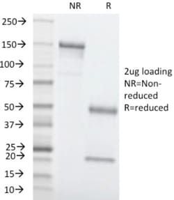
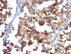
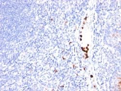
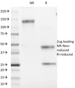
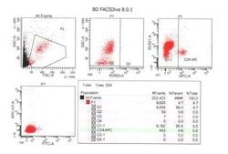
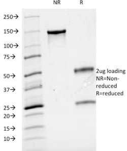
Flow Cytometry, Immunohistochemistry (Paraffin), SDS-Page, Immunofluorescence
Conjugate
Unconjugated
Host Species
Mouse
Research Discipline
Dendritic Cell Markers, Immunology
Formulation
10mM PBS and 0.05% BSA with 0.05% Sodium Azide
Gene ID (Entrez)
909
Immunogen
Human thymocytes
Primary or Secondary
Primary
Content And Storage
Store at 4C.
Molecular Weight of Antigen
49 kDa
Clone
66IIC7
Dilution
Flow Cytometry 0.5 - 1 ug/million cells in 0.1 ml, Immunohistochemistry-Paraffin 0.5 - 1.0 ug/ml, SDS-Page, Immunofluorescence 1 - 2 ug/ml
Classification
Monoclonal
Form
Purified
Regulatory Status
RUO
Target Species
Human
Gene Alias
CD1, CD1a antigen, CD1A antigen, a polypeptide, CD1a molecule, cluster of differentiation 1 A, cortical thymocyte antigen CD1A, differentiation antigen CD1-alpha-3, epidermal dendritic cell marker CD1a, FCB6, HTA1, hTa1 thymocyte antigen, R4, T6, T-cell surface antigen T6/Leu-6, T-cell surface glycoprotein CD1a
Gene Symbols
CD1A
Isotype
IgG2a κ
Purification Method
Protein A or G purified
Test Specificity
At least five CD1 genes (CD1a, b, c, d, and e) are identified. CD1 proteins have been demonstrated to restrict T cell response to non-peptide lipid and glycolipid antigens and play a role in non-classical antigen presentation. CD1a is a non-polymorphic MHC Class 1 related cell surface glycoprotein, expressed in association with Beta-2 microglobulin. Anti-CD1a labels Langerhans cell histiocytosis (Histiocytosis X), extranodal histiocytic sarcoma, a subset of T-lymphoblastic lymphoma/leukemia, and interdigitating dendritic cell sarcoma of the lymph node. When combined with antibodies against TTF-1 and CD5, anti-CD1a is useful in distinguishing between pulmonary and thymic neoplasms since CD1a is consistently expressed in thymic lymphocytes in both typical and atypical thymomas, but only focally in 1/6 of thymic carcinomas and not in lymphocytes in pulmonary neoplasms. Anti-CD1a is reported to be a new marker for perivascular epithelial cell tumor (PEComa)
Related Products


Description
- Ensure accurate, reproducible results in Flow Cytometry, Immunohistochemistry (Paraffin), Immunofluorescence CD1a Monoclonal specifically detects CD1a in Human samples
- It is validated for Flow Cytometry, Immunocytochemistry/Immunofluorescence, Immunofluorescence.
Compare Similar Items
Show Difference
Antigen: CD1a
Concentration: 0.2 mg/mL
Applications: Flow Cytometry, Immunohistochemistry (Paraffin), SDS-Page, Immunofluorescence
Conjugate: Unconjugated
Host Species: Mouse
Research Discipline: Dendritic Cell Markers, Immunology
Formulation: 10mM PBS and 0.05% BSA with 0.05% Sodium Azide
Gene ID (Entrez): 909
Immunogen: Human thymocytes
Primary or Secondary: Primary
Content And Storage: Store at 4C.
Molecular Weight of Antigen: 49 kDa
Clone: 66IIC7
Dilution: Flow Cytometry 0.5 - 1 ug/million cells in 0.1 ml, Immunohistochemistry-Paraffin 0.5 - 1.0 ug/ml, SDS-Page, Immunofluorescence 1 - 2 ug/ml
Classification: Monoclonal
Form: Purified
Regulatory Status: RUO
Target Species: Human
Gene Alias: CD1, CD1a antigen, CD1A antigen, a polypeptide, CD1a molecule, cluster of differentiation 1 A, cortical thymocyte antigen CD1A, differentiation antigen CD1-alpha-3, epidermal dendritic cell marker CD1a, FCB6, HTA1, hTa1 thymocyte antigen, R4, T6, T-cell surface antigen T6/Leu-6, T-cell surface glycoprotein CD1a
Gene Symbols: CD1A
Isotype: IgG2a κ
Purification Method: Protein A or G purified
Test Specificity: At least five CD1 genes (CD1a, b, c, d, and e) are identified. CD1 proteins have been demonstrated to restrict T cell response to non-peptide lipid and glycolipid antigens and play a role in non-classical antigen presentation. CD1a is a non-polymorphic MHC Class 1 related cell surface glycoprotein, expressed in association with Beta-2 microglobulin. Anti-CD1a labels Langerhans cell histiocytosis (Histiocytosis X), extranodal histiocytic sarcoma, a subset of T-lymphoblastic lymphoma/leukemia, and interdigitating dendritic cell sarcoma of the lymph node. When combined with antibodies against TTF-1 and CD5, anti-CD1a is useful in distinguishing between pulmonary and thymic neoplasms since CD1a is consistently expressed in thymic lymphocytes in both typical and atypical thymomas, but only focally in 1/6 of thymic carcinomas and not in lymphocytes in pulmonary neoplasms. Anti-CD1a is reported to be a new marker for perivascular epithelial cell tumor (PEComa)
Antigen: CD2
Concentration: 0.2 mg/mL
Applications: Flow Cytometry, Immunofluorescence
Conjugate: Unconjugated
Host Species: Mouse
Research Discipline: Adaptive Immunity, Apoptosis, Immunology
Formulation: 10mM PBS and 0.05% BSA with 0.05% Sodium Azide
Gene ID (Entrez): 914
Immunogen: Human thymocytes
Primary or Secondary: Primary
Content And Storage: Store at 4C.
Molecular Weight of Antigen: 50 kDa
Clone: HuLy-m1
Dilution: Flow Cytometry 0.5 - 1 ug/million cells in 0.1 ml, Immunofluorescence 0.5 - 1.0 ug/ml
Classification: Monoclonal
Form: Purified
Regulatory Status: RUO
Target Species: Human, Feline
Gene Alias: CD2 antigen, CD2 antigen (p50), sheep red blood cell receptor, CD2 molecule, Erythrocyte receptor, FLJ46032, LFA-2, LFA-3 receptor, lymphocyte-function antigen-2, Rosette receptor, SRBC, T11, T-cell surface antigen CD2, T-cell surface antigen T11/Leu-5
Gene Symbols: CD2
Isotype: IgG2b κ
Purification Method: Protein A or G purified
Test Specificity: CD2 interacts through its amino-terminal domain with the extracellular domain of CD58 (also designated CD2 ligand) to mediate cell adhesion. CD2/CD58 binding can enhance antigen-specific T cell activation. CD2 is a transmembrane glycoprotein that is expressed on peripheral blood T lymphocytes, NK cells and thymocytes. CD58 is a heavily glycosylated protein with a broad tissue distribution in hematopoietic and other cells, including endothelium. Interaction between CD2 and its counter receptor LFA3 (CD58) on opposing cells optimizes immune system recognition, thereby facilitating communication between helper T lymphocytes and antigen-presenting cells, as well as between cytolytic effectors and target cells.
Antigen: CD3 epsilon
Concentration: 0.2 mg/mL
Applications: Flow Cytometry, SDS-Page, Immunofluorescence
Conjugate: Unconjugated
Host Species: Mouse
Research Discipline: Adaptive Immunity, Apoptosis, Cytokine Research, Diabetes Research, Immunology, Innate Immunity, Mesenchymal Stem Cell Markers, Signal Transduction, Stem Cell Lines, Stem Cell Markers
Formulation: 10mM PBS and 0.05% BSA with 0.05% Sodium Azide
Gene ID (Entrez): 916
Immunogen: Human T cell leukemia cells
Primary or Secondary: Primary
Content And Storage: Store at 4C.
Molecular Weight of Antigen: 20 kDa
Clone: B-B12
Dilution: Flow Cytometry 0.5 - 1 ug/million cells in 0.1 ml, SDS-Page, Immunofluorescence 1 - 2 ug/ml
Classification: Monoclonal
Form: Purified
Regulatory Status: RUO
Target Species: Human
Gene Alias: CD3e antigen, CD3e antigen, epsilon polypeptide (TiT3 complex), CD3e molecule, epsilon (CD3-TCR complex), CD3-epsilon, FLJ18683, T3E, T-cell antigen receptor complex, epsilon subunit of T3, T-cell surface antigen T3/Leu-4 epsilon chain, T-cell surface glycoprotein CD3 epsilon chain, TCRE
Gene Symbols: CD3E
Isotype: IgG1 κ
Purification Method: Protein A purified
Test Specificity: Reacts with five invariable CD3 chains (designated as and ) with molecular weight ranging from 16-28kDa. CD3 is expressed, typically at high levels, on peripheral T cells and majority of T cell neoplasms. Thymocytes express CD3 at different level on the cell surface in the course of differentiation and, in cortical thymus, CD3 is predominantly Intracytoplasmic. The CD3 complex is closely associated at the lymphocyte cell surface with T cell antigen receptor (TCR) and is involved in transducing antigen-recognition signals into cytoplasm of T cells and in regulating the cell surface expression of the TCR complex.