CEACAM5/CD66e Antibody (SPM506), Novus Biologicals™
Mouse Monoclonal Antibody
Manufacturer: Fischer Scientific
The price for this product is unavailable. Please request a quote
Antigen
CEACAM5/CD66e
Concentration
0.2mg/mL
Applications
Flow Cytometry, Immunohistochemistry (Paraffin), Immunofluorescence
Conjugate
Unconjugated
Host Species
Mouse
Research Discipline
Cancer, Cellular Markers, Immunology
Formulation
10mM PBS and 0.05% BSA with 0.05% Sodium Azide
Gene Alias
Carcinoembryonic antigen, carcinoembryonic antigen-related cell adhesion molecule 5, CD66e antigen, CEACD66e, DKFZp781M2392, Meconium antigen 100
Gene Symbols
CEACAM5
Isotype
IgG1 κ
Purification Method
Protein A or G purified
Test Specificity
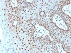
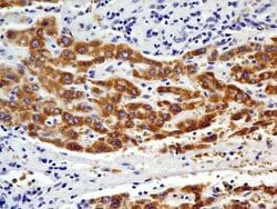
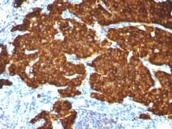
This antibody recognizes proteins of 80-200kDa, identified as different members of CEA family. CEA is synthesized during development in the fetal gut and is re-expressed in increased amounts in intestinal carcinomas and several other tumors. This MAb does not react with nonspecific cross-reacting antigen (NCA) and with human polymorphonuclear leucocytes. It shows no reaction with a variety of normal tissues and is suitable for staining of formalin/paraffin tissues. CEA is not found in benign glands, stroma, or malignant prostatic cells. Antibody to CEA is useful in detecting early foci of gastric carcinoma and in distinguishing pulmonary adenocarcinomas (60-70% are CEA+) from pleural mesotheliomas (rarely or weakly CEA+). Anti-CEA positivity is seen in adenocarcinomas from the lung, colon, stomach, esophagus, pancreas, gallbadder, urachus, salivary gland, ovary, and endocervix.
Clone
SPM506
Dilution
Flow Cytometry 0.5 - 1 ug/million cells in 0.1 ml, Immunohistochemistry-Paraffin 0.25 - 0.5 ug/ml, Immunofluorescence 1 - 2 ug/ml
Classification
Monoclonal
Form
Purified
Regulatory Status
RUO
Target Species
Human, Monkey
Gene Accession No.
P06731
Gene ID (Entrez)
1048
Immunogen
Human colon carcinoma extract
Primary or Secondary
Primary
Content And Storage
Store at 4C.
Description
- Ensure accurate, reproducible results in Flow Cytometry, Immunohistochemistry (Paraffin), Immunofluorescence CEACAM5/CD66e Monoclonal specifically detects CEACAM5/CD66e in Human, Monkey samples
- It is validated for Flow Cytometry, Immunohistochemistry, Immunocytochemistry/Immunofluorescence, Immunohistochemistry-Paraffin, Immunofluorescence.
Compare Similar Items
Show Difference

Antigen: CEACAM5/CD66e
Concentration: 0.2mg/mL
Applications: Flow Cytometry, Immunohistochemistry (Paraffin), Immunofluorescence
Conjugate: Unconjugated
Host Species: Mouse
Research Discipline: Cancer, Cellular Markers, Immunology
Formulation: 10mM PBS and 0.05% BSA with 0.05% Sodium Azide
Gene Alias: Carcinoembryonic antigen, carcinoembryonic antigen-related cell adhesion molecule 5, CD66e antigen, CEACD66e, DKFZp781M2392, Meconium antigen 100
Gene Symbols: CEACAM5
Isotype: IgG1 κ
Purification Method: Protein A or G purified
Test Specificity: This antibody recognizes proteins of 80-200kDa, identified as different members of CEA family. CEA is synthesized during development in the fetal gut and is re-expressed in increased amounts in intestinal carcinomas and several other tumors. This MAb does not react with nonspecific cross-reacting antigen (NCA) and with human polymorphonuclear leucocytes. It shows no reaction with a variety of normal tissues and is suitable for staining of formalin/paraffin tissues. CEA is not found in benign glands, stroma, or malignant prostatic cells. Antibody to CEA is useful in detecting early foci of gastric carcinoma and in distinguishing pulmonary adenocarcinomas (60-70% are CEA+) from pleural mesotheliomas (rarely or weakly CEA+). Anti-CEA positivity is seen in adenocarcinomas from the lung, colon, stomach, esophagus, pancreas, gallbadder, urachus, salivary gland, ovary, and endocervix.
Clone: SPM506
Dilution: Flow Cytometry 0.5 - 1 ug/million cells in 0.1 ml, Immunohistochemistry-Paraffin 0.25 - 0.5 ug/ml, Immunofluorescence 1 - 2 ug/ml
Classification: Monoclonal
Form: Purified
Regulatory Status: RUO
Target Species: Human, Monkey
Gene Accession No.: P06731
Gene ID (Entrez): 1048
Immunogen: Human colon carcinoma extract
Primary or Secondary: Primary
Content And Storage: Store at 4C.
Antigen: CHREBP
Concentration: 1.0 mg/mL
Applications: Western Blot, ChIP Assay, Immunohistochemistry, Immunocytochemistry, Immunofluorescence
Conjugate: Unconjugated
Host Species: Mouse
Research Discipline: Cancer, Chromatin Research, DNA replication Transcription Translation and Splicing, Lipid and Metabolism, Transcription Factors and Regulators
Formulation: PBS with 0.02% Sodium Azide
Gene Alias: BHLHD14, CHREBPcarbohydrate-responsive element-binding protein, Class D basic helix-loop-helix protein 14, MIOWS basic-helix-loop-helix leucine zipper protein, MLX interacting protein-like, MLX interactor, MLX-interacting protein-like, MONDOBWilliams-Beuren syndrome chromosomal region 14 protein, WBSCR14Williams-Beuren syndrome chromosome region 14 protein 1, Williams Beuren syndrome chromosome region 14, Williams-Beuren syndrome chromosome region 14 protein 2, WS-bHLHbHLHd14carbohydrate response element binding protein
Gene Symbols: MLXIPL
Isotype: IgG2b κ
Purification Method: Protein G purified
Test Specificity: __
Clone: 2D9NB
Dilution: Western Blot 1:500 ug/ml, Chromatin Immunoprecipitation reported in scientific literature (PMID 31289120), Immunohistochemistry 1:200, Immunocytochemistry/Immunofluorescence 1:100, Immunohistochemistry-Paraffin 1:200, Chromatin Immunoprecipitation (ChIP)
Classification: Monoclonal
Form: Purified
Regulatory Status: RUO
Target Species: Human, Mouse, Rat
Gene Accession No.: Q9NP71-1
Gene ID (Entrez): 51085
Immunogen: Partial recombinant human ChREBP protein between amino acids 600-800. [Uniprot: Q9NP71]
Primary or Secondary: Primary
Content And Storage: Store at 4C short term. Aliquot and store at -20C long term. Avoid freeze-thaw cycles.
Antigen: Chromogranin A
Concentration: 0.2mg/mL
Applications: Flow Cytometry, Immunohistochemistry (Paraffin), Immunofluorescence
Conjugate: Unconjugated
Host Species: Mouse
Research Discipline: Apoptosis, Cancer, Neuronal Cell Markers, Tumor Biomarkers
Formulation: 10mM PBS and 0.05% BSA with 0.05% Sodium Azide
Gene Alias: betagranin (N-terminal fragment of chromogranin A), CGA, chromogranin A (parathyroid secretory protein 1), chromogranin-A, parathyroid secretory protein 1, Pituitary secretory protein I, SP-I
Gene Symbols: CHGA
Isotype: IgG
Purification Method: Protein A or G purified
Test Specificity: Chromogranin A is present in neuroendocrine cells throughout the body, including the neuroendocrine cells of the large and small intestine, adrenal medulla and pancreatic islets. It is an excellent marker for carcinoid tumors, pheochromocytomas, paragangliomas, and other neuroendocrine tumors. Co-expression of chromogranin A and neuron specific enolase (NSE) is common in neuroendocrine neoplasms. Reportedly, co-expression of certain keratins and chromogranin indicates neuroendocrine lineage. The presence of strong anti-chromogranin staining and absence of anti-keratin staining should raise the possibility of paraganglioma. The co-expression of chromogranin and NSE is typical of neuroendocrine neoplasms. Most pituitary adenomas and prolactinomas readily express chromogranin.
Clone: CGA/413 + CHGA/777 + CHGA/798
Dilution: Flow Cytometry 0.5 - 1 ug/million cells in 0.1 ml, Immunohistochemistry-Paraffin 0.25 - 0.5 ug/ml, Immunofluorescence 0.5 - 1.0 ug/ml
Classification: Monoclonal
Form: Purified
Regulatory Status: RUO
Target Species: Human, Mouse, Rat, Porcine, Monkey
Gene Accession No.: P10645
Gene ID (Entrez): 1113
Immunogen: Recombinant human chromogranin A protein
Primary or Secondary: Primary
Content And Storage: Store at 4C.