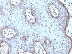
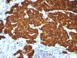
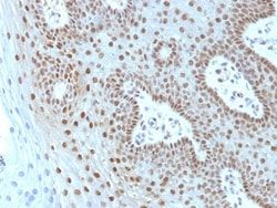
Chromogranin A Antibody (CGA/413 + CHGA/777 + CHGA/798), Novus Biologicals™
Mouse Monoclonal Antibody has been used in 1 publication
Manufacturer: Fischer Scientific
The price for this product is unavailable. Please request a quote
Antigen
Chromogranin A
Concentration
0.2mg/mL
Applications
Flow Cytometry, Immunohistochemistry (Paraffin), Immunofluorescence
Conjugate
Unconjugated
Host Species
Mouse
Research Discipline
Apoptosis, Cancer, Neuronal Cell Markers, Tumor Biomarkers
Formulation
10mM PBS and 0.05% BSA with 0.05% Sodium Azide
Gene Alias
betagranin (N-terminal fragment of chromogranin A), CGA, chromogranin A (parathyroid secretory protein 1), chromogranin-A, parathyroid secretory protein 1, Pituitary secretory protein I, SP-I
Gene Symbols
CHGA
Isotype
IgG
Purification Method
Protein A or G purified
Test Specificity
Chromogranin A is present in neuroendocrine cells throughout the body, including the neuroendocrine cells of the large and small intestine, adrenal medulla and pancreatic islets. It is an excellent marker for carcinoid tumors, pheochromocytomas, paragangliomas, and other neuroendocrine tumors. Co-expression of chromogranin A and neuron specific enolase (NSE) is common in neuroendocrine neoplasms. Reportedly, co-expression of certain keratins and chromogranin indicates neuroendocrine lineage. The presence of strong anti-chromogranin staining and absence of anti-keratin staining should raise the possibility of paraganglioma. The co-expression of chromogranin and NSE is typical of neuroendocrine neoplasms. Most pituitary adenomas and prolactinomas readily express chromogranin.
Clone
CGA/413 + CHGA/777 + CHGA/798
Dilution
Flow Cytometry 0.5 - 1 ug/million cells in 0.1 ml, Immunohistochemistry-Paraffin 0.25 - 0.5 ug/ml, Immunofluorescence 0.5 - 1.0 ug/ml
Classification
Monoclonal
Form
Purified
Regulatory Status
RUO
Target Species
Human, Mouse, Rat, Porcine, Monkey
Gene Accession No.
P10645
Gene ID (Entrez)
1113
Immunogen
Recombinant human chromogranin A protein
Primary or Secondary
Primary
Content And Storage
Store at 4C.
Description
- Ensure accurate, reproducible results in Flow Cytometry, Immunohistochemistry (Paraffin), Immunofluorescence Chromogranin A Monoclonal specifically detects Chromogranin A in Human, Mouse, Rat, Porcine, Monkey samples
- It is validated for Western Blot, Immunohistochemistry, Immunocytochemistry/Immunofluorescence, Immunohistochemistry-Paraffin.
Compare Similar Items
Show Difference
Antigen: Chromogranin A
Concentration: 0.2mg/mL
Applications: Flow Cytometry, Immunohistochemistry (Paraffin), Immunofluorescence
Conjugate: Unconjugated
Host Species: Mouse
Research Discipline: Apoptosis, Cancer, Neuronal Cell Markers, Tumor Biomarkers
Formulation: 10mM PBS and 0.05% BSA with 0.05% Sodium Azide
Gene Alias: betagranin (N-terminal fragment of chromogranin A), CGA, chromogranin A (parathyroid secretory protein 1), chromogranin-A, parathyroid secretory protein 1, Pituitary secretory protein I, SP-I
Gene Symbols: CHGA
Isotype: IgG
Purification Method: Protein A or G purified
Test Specificity: Chromogranin A is present in neuroendocrine cells throughout the body, including the neuroendocrine cells of the large and small intestine, adrenal medulla and pancreatic islets. It is an excellent marker for carcinoid tumors, pheochromocytomas, paragangliomas, and other neuroendocrine tumors. Co-expression of chromogranin A and neuron specific enolase (NSE) is common in neuroendocrine neoplasms. Reportedly, co-expression of certain keratins and chromogranin indicates neuroendocrine lineage. The presence of strong anti-chromogranin staining and absence of anti-keratin staining should raise the possibility of paraganglioma. The co-expression of chromogranin and NSE is typical of neuroendocrine neoplasms. Most pituitary adenomas and prolactinomas readily express chromogranin.
Clone: CGA/413 + CHGA/777 + CHGA/798
Dilution: Flow Cytometry 0.5 - 1 ug/million cells in 0.1 ml, Immunohistochemistry-Paraffin 0.25 - 0.5 ug/ml, Immunofluorescence 0.5 - 1.0 ug/ml
Classification: Monoclonal
Form: Purified
Regulatory Status: RUO
Target Species: Human, Mouse, Rat, Porcine, Monkey
Gene Accession No.: P10645
Gene ID (Entrez): 1113
Immunogen: Recombinant human chromogranin A protein
Primary or Secondary: Primary
Content And Storage: Store at 4C.
Antigen: c-Myc
Concentration: 0.2 mg/mL
Applications: Flow Cytometry, Immunohistochemistry (Paraffin), Immunofluorescence
Conjugate: Unconjugated
Host Species: Mouse
Research Discipline: Autophagy, Cancer, Cancer Stem Cells, Cell Cycle and Replication, Chromatin Research, Core ESC Like Genes, Epitope Tags, Myc Epitope Tags, Stem Cell Markers, Transcription Factors and Regulators, Tumor Suppressors
Formulation: 10mM PBS and 0.05% BSA with 0.05% Sodium Azide
Gene Alias: avian myelocytomatosis viral oncogene homolog, BHLHE39, bHLHe39MRTL, Class E basic helix-loop-helix protein 39, c-Myc, MYC, myc proto-oncogene protein, MYCC, myc-related translation/localization regulatory factor, Proto-oncogene c-Myc, Transcription factor p64, v-myc avian myelocytomatosis viral oncogene homolog, v-myc myelocytomatosis viral oncogene homolog (avian)
Gene Symbols: MYC
Isotype: IgG1 κ
Purification Method: Protein A or G purified
Test Specificity: It recognizes a transcription factor of 64-67kDa, identified as c-myc. This MAb shows no cross-reaction with v-myc. c-myc is involved in the control of cell proliferation and differentiation and is amplified and/or over-expressed in a variety of tumors. Over-expression of c-myc protein occurs frequently in luminal cells of prostate intraepithelial neoplasia as well as in most primary carcinomas and metastatic disease. Rearrangement of the MYC gene is found in 3% to 16% of diffuse large B-cell lymphoma (DLBCL s) and in nearly 100% of Burkitt lymphomas (BL). Identifying MYC status is important in establishing final diagnosis of DLBCL, BL, or B-cell lymphoma, with features intermediate between DLBCL and BL as well as in differential diagnoses of the lymphomas.
Clone: MYC275 + MYC909
Dilution: Flow Cytometry 0.5 - 1 ug/million cells, Immunohistochemistry-Paraffin 1 - 2 ug/ml, Immunofluorescence 1 - 2 ug/ml
Classification: Monoclonal
Form: Purified
Regulatory Status: RUO
Target Species: Human
Gene Accession No.: P01106
Gene ID (Entrez): 4609
Immunogen: Recombinant human c-myc protein
Primary or Secondary: Primary
Content And Storage: Store at 4C.