c-Myc Antibody (MYC909), Novus Biologicals™
Mouse Monoclonal Antibody
Manufacturer: Fischer Scientific
The price for this product is unavailable. Please request a quote
Antigen
c-Myc
Concentration
0.2mg/mL
Applications
Flow Cytometry, Immunohistochemistry (Paraffin), Immunofluorescence
Conjugate
Unconjugated
Host Species
Mouse
Research Discipline
Autophagy, Cancer, Cancer Stem Cells, Cell Cycle and Replication, Chromatin Research, Core ESC Like Genes, Epitope Tags, Myc Epitope Tags, Stem Cell Markers, Transcription Factors and Regulators, Tumor Suppressors
Formulation
10mM PBS and 0.05% BSA with 0.05% Sodium Azide
Gene ID (Entrez)
4609
Immunogen
Recombinant human c-myc protein
Primary or Secondary
Primary
Content And Storage
Store at 4C.
Clone
MYC909
Dilution
Flow Cytometry 0.5 - 1 ug/million cells in 0.1 ml, Immunohistochemistry-Paraffin 1 - 2 ug/ml, Immunofluorescence 1 - 2 ug/ml
Classification
Monoclonal
Form
Purified
Regulatory Status
RUO
Target Species
Human
Gene Alias
avian myelocytomatosis viral oncogene homolog, BHLHE39, bHLHe39MRTL, Class E basic helix-loop-helix protein 39, c-Myc, MYC, myc proto-oncogene protein, MYCC, myc-related translation/localization regulatory factor, Proto-oncogene c-Myc, Transcription factor p64, v-myc avian myelocytomatosis viral oncogene homolog, v-myc myelocytomatosis viral oncogene homolog (avian)
Gene Symbols
MYC
Isotype
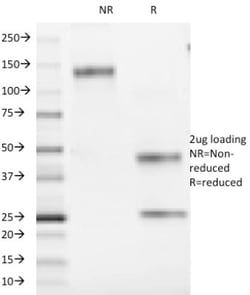
IgG1 κ
Purification Method
Protein A or G purified
Test Specificity
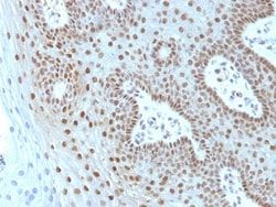
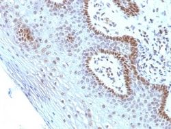
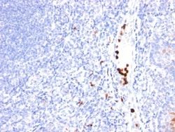
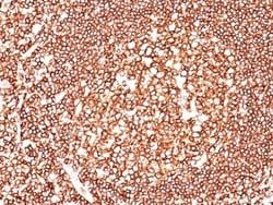
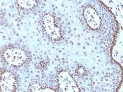
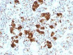
It recognizes a transcription factor of 64-67kDa, identified as c-myc. This MAb shows no cross-reaction with v-myc. c-myc is involved in the control of cell proliferation and differentiation and is amplified and/or over-expressed in a variety of tumors. Over-expression of c-myc protein occurs frequently in luminal cells of prostate intraepithelial neoplasia as well as in most primary carcinomas and metastatic disease. Rearrangement of the MYC gene is found in 3% to 16% of diffuse large B-cell lymphoma (DLBCLs) and in nearly 100% of Burkitt lymphomas (BL). Identifying MYC status is important in establishing final diagnosis of DLBCL, BL, or B-cell lymphoma, with features intermediate between DLBCL and BL as well as in differential diagnoses of the lymphomas.
Related Products

Description
- Ensure accurate, reproducible results in Flow Cytometry, Immunohistochemistry (Paraffin), Immunofluorescence c-Myc Monoclonal specifically detects c-Myc in Human samples
- It is validated for Flow Cytometry, Immunohistochemistry, Immunocytochemistry/Immunofluorescence, Immunohistochemistry-Paraffin, Immunofluorescence.
Compare Similar Items
Show Difference
Antigen: c-Myc
Concentration: 0.2mg/mL
Applications: Flow Cytometry, Immunohistochemistry (Paraffin), Immunofluorescence
Conjugate: Unconjugated
Host Species: Mouse
Research Discipline: Autophagy, Cancer, Cancer Stem Cells, Cell Cycle and Replication, Chromatin Research, Core ESC Like Genes, Epitope Tags, Myc Epitope Tags, Stem Cell Markers, Transcription Factors and Regulators, Tumor Suppressors
Formulation: 10mM PBS and 0.05% BSA with 0.05% Sodium Azide
Gene ID (Entrez): 4609
Immunogen: Recombinant human c-myc protein
Primary or Secondary: Primary
Content And Storage: Store at 4C.
Clone: MYC909
Dilution: Flow Cytometry 0.5 - 1 ug/million cells in 0.1 ml, Immunohistochemistry-Paraffin 1 - 2 ug/ml, Immunofluorescence 1 - 2 ug/ml
Classification: Monoclonal
Form: Purified
Regulatory Status: RUO
Target Species: Human
Gene Alias: avian myelocytomatosis viral oncogene homolog, BHLHE39, bHLHe39MRTL, Class E basic helix-loop-helix protein 39, c-Myc, MYC, myc proto-oncogene protein, MYCC, myc-related translation/localization regulatory factor, Proto-oncogene c-Myc, Transcription factor p64, v-myc avian myelocytomatosis viral oncogene homolog, v-myc myelocytomatosis viral oncogene homolog (avian)
Gene Symbols: MYC
Isotype: IgG1 κ
Purification Method: Protein A or G purified
Test Specificity: It recognizes a transcription factor of 64-67kDa, identified as c-myc. This MAb shows no cross-reaction with v-myc. c-myc is involved in the control of cell proliferation and differentiation and is amplified and/or over-expressed in a variety of tumors. Over-expression of c-myc protein occurs frequently in luminal cells of prostate intraepithelial neoplasia as well as in most primary carcinomas and metastatic disease. Rearrangement of the MYC gene is found in 3% to 16% of diffuse large B-cell lymphoma (DLBCLs) and in nearly 100% of Burkitt lymphomas (BL). Identifying MYC status is important in establishing final diagnosis of DLBCL, BL, or B-cell lymphoma, with features intermediate between DLBCL and BL as well as in differential diagnoses of the lymphomas.
Antigen: c-Myc
Concentration: 0.2mg/mL
Applications: Flow Cytometry, Immunohistochemistry (Paraffin), Immunofluorescence
Conjugate: Unconjugated
Host Species: Mouse
Research Discipline: Autophagy, Cancer, Cancer Stem Cells, Cell Cycle and Replication, Chromatin Research, Core ESC Like Genes, Epitope Tags, Myc Epitope Tags, Stem Cell Markers, Transcription Factors and Regulators, Tumor Suppressors
Formulation: 10mM PBS and 0.05% BSA with 0.05% Sodium Azide
Gene ID (Entrez): 4609
Immunogen: A synthetic peptide, corresponding to aa 408-439 (AEEQKLISEEDLLRKRREQLKHKLEQLRNSCA) from C-terminus of human c-myc, coupled to KLH.
Primary or Secondary: Primary
Content And Storage: Store at 4C.
Clone: SPM237
Dilution: Flow Cytometry 0.5 - 1 ug/million cells in 0.1 ml, Immunohistochemistry-Paraffin 1 - 2 ug/ml, Immunofluorescence 1 - 2 ug/ml
Classification: Monoclonal
Form: Purified
Regulatory Status: RUO
Target Species: Human, Chicken (Negative), Mouse (Negative), Rat (Negative)
Gene Alias: avian myelocytomatosis viral oncogene homolog, BHLHE39, bHLHe39MRTL, Class E basic helix-loop-helix protein 39, c-Myc, MYC, myc proto-oncogene protein, MYCC, myc-related translation/localization regulatory factor, Proto-oncogene c-Myc, Transcription factor p64, v-myc avian myelocytomatosis viral oncogene homolog, v-myc myelocytomatosis viral oncogene homolog (avian)
Gene Symbols: MYC
Isotype: IgG1 κ
Purification Method: Protein A or G purified
Test Specificity: It recognizes a transcription factor of 64-67kDa, identified as c-myc. Its epitope spans between aa 410-419 (EQKLISEEDL) which is a specific portion of an alpha helical region of human c-myc protein. This MAb shows no cross-reaction with v-myc. c-myc is involved in the control of cell proliferation and differentiation and is amplified and/or overexpressed in a variety of tumors. Over-expression of c-myc protein occurs frequently in luminal cells of prostate intraepithelial neoplasia as well as in most primary carcinomas and metastatic disease
Antigen: CPS1
Concentration: 0.2mg/mL
Applications: Immunohistochemistry (Paraffin), Immunofluorescence
Conjugate: Unconjugated
Host Species: Mouse
Research Discipline: __
Formulation: 10mM PBS and 0.05% BSA with 0.05% Sodium Azide
Gene ID (Entrez): 1373
Immunogen: Recombinant human CPS1 protein
Primary or Secondary: Primary
Content And Storage: Store at 4C.
Clone: CPS1/1022
Dilution: Immunohistochemistry-Paraffin 0.25 - 0.5 ug/ml, Immunofluorescence 0.5 - 1.0 ug/ml
Classification: Monoclonal
Form: Purified
Regulatory Status: RUO
Target Species: Human, Canine
Gene Alias: carbamoyl-phosphate synthase [ammonia], mitochondrial, carbamoyl-phosphate synthase 1, mitochondrial, carbamoyl-phosphate synthetase 1, mitochondrial, carbamoylphosphate synthetase I, Carbamoyl-phosphate synthetase I, CPSase I, CPSASE1, EC 6.3.4.16
Gene Symbols: CPS1
Isotype: IgG1
Purification Method: Protein A or G purified
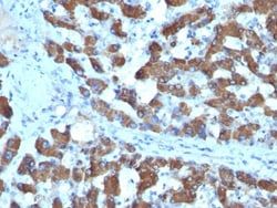
Test Specificity: This MAb recognizes a protein of 165kDa, identified as carbamoyl phosphate synthetase 1 (CPS1). This mitochondrial enzyme catalyzes synthesis of carbamoyl phosphate from ammonia and bicarbonate. This reaction is the first committed step of the urea cycle, which is important in the removal of excess urea from cells.Deficiency of CPS1 is an autosomal recessive disorder that causes hyperammonemia. CPS1 is a hepatocyte specific protein that localizes to the mitochondria of hepatocytes. It is a sensitive marker for distinguishing hepatocellular carcinomas (HCC) from other metastatic carcinomas as well as cholangio-carcinomas. HCC s occur primarily in the stomach, but they are also found in many other organs. CPS1 may also be a useful marker for intestinal metaplasia. Reportedly, strong expression of CPS1 correlates with smaller tumor size and longer patient survival. Occasionally, CPS1 is also found in gastric carcinomas as well as in a few other non-hepatic tumors.