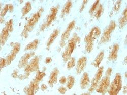
Cytokeratin 8/18 Antibody (SPM141), Novus Biologicals™
Mouse Monoclonal Antibody
Manufacturer: Fischer Scientific
The price for this product is unavailable. Please request a quote
Antigen
Cytokeratin 8/18
Concentration
0.2mg/mL
Applications
Western Blot, Flow Cytometry, Immunohistochemistry (Paraffin), Immunofluorescence
Conjugate
Unconjugated
Host Species
Mouse
Research Discipline
Cancer, Cytoskeleton Markers
Formulation
10mM PBS and 0.05% BSA with 0.05% Sodium Azide
Gene ID (Entrez)
3856
Immunogen
Keratin preparation from human breast carcinoma MCF-7 cells
Primary or Secondary
Primary
Content And Storage
Store at 4C.
Clone
SPM141
Dilution
Western Blot 0.5 - 1.0 ug/ml, Flow Cytometry 0.5 - 1 ug/million cells in 0.1 ml, Immunohistochemistry-Paraffin 0.5 - 1.0 ug/ml, Immunofluorescence 1 - 2 ug/ml
Classification
Monoclonal
Form
Purified
Regulatory Status
RUO
Target Species
Human
Gene Alias
CARD2, CK8, CK-8, CYK8cytokeratin 8, Cytokeratin-8, K2C8, K8cytokeratin-8, keratin 8, keratin, type II cytoskeletal 8, keratin-8, KO, Type-II keratin Kb8
Gene Symbols
KRT8
Isotype
IgG1 κ
Purification Method
Protein A or G purified
Test Specificity
Cytokeratin 8 (CK8) belongs to the type II (or B or basic) subfamily of high molecular weight cytokeratins and exists in combination with cytokeratin 18 (CK18). This MAb cocktail recognizes all simple epithelia including glandular epithelium, for example thyroid, female breast, gastrointestinal tract, respiratory tract, and urogenital tract including transitional epithelium. All adenocarcinomas and most squamous carcinomas are positive but keratinizing squamous carcinomas are usually negative. This antibody is useful in demonstrating the presence of Paget cells; there is very little keratin 18 in the normal epidermis so only Paget cells are stained.
Description
- Ensure accurate, reproducible results in Western Blot, Flow Cytometry, Immunohistochemistry (Paraffin), Immunofluorescence Cytokeratin 8/18 Monoclonal specifically detects Cytokeratin 8/18 in Human samples
- It is validated for Western Blot, Flow Cytometry, Immunohistochemistry, Immunocytochemistry/Immunofluorescence, Immunohistochemistry-Paraffin, Immunofluorescence.
Compare Similar Items
Show Difference

Antigen: Cytokeratin 8/18
Concentration: 0.2mg/mL
Applications: Western Blot, Flow Cytometry, Immunohistochemistry (Paraffin), Immunofluorescence
Conjugate: Unconjugated
Host Species: Mouse
Research Discipline: Cancer, Cytoskeleton Markers
Formulation: 10mM PBS and 0.05% BSA with 0.05% Sodium Azide
Gene ID (Entrez): 3856
Immunogen: Keratin preparation from human breast carcinoma MCF-7 cells
Primary or Secondary: Primary
Content And Storage: Store at 4C.
Clone: SPM141
Dilution: Western Blot 0.5 - 1.0 ug/ml, Flow Cytometry 0.5 - 1 ug/million cells in 0.1 ml, Immunohistochemistry-Paraffin 0.5 - 1.0 ug/ml, Immunofluorescence 1 - 2 ug/ml
Classification: Monoclonal
Form: Purified
Regulatory Status: RUO
Target Species: Human
Gene Alias: CARD2, CK8, CK-8, CYK8cytokeratin 8, Cytokeratin-8, K2C8, K8cytokeratin-8, keratin 8, keratin, type II cytoskeletal 8, keratin-8, KO, Type-II keratin Kb8
Gene Symbols: KRT8
Isotype: IgG1 κ
Purification Method: Protein A or G purified
Test Specificity: Cytokeratin 8 (CK8) belongs to the type II (or B or basic) subfamily of high molecular weight cytokeratins and exists in combination with cytokeratin 18 (CK18). This MAb cocktail recognizes all simple epithelia including glandular epithelium, for example thyroid, female breast, gastrointestinal tract, respiratory tract, and urogenital tract including transitional epithelium. All adenocarcinomas and most squamous carcinomas are positive but keratinizing squamous carcinomas are usually negative. This antibody is useful in demonstrating the presence of Paget cells; there is very little keratin 18 in the normal epidermis so only Paget cells are stained.
Antigen: Cytokeratin, LMW
Concentration: 0.2mg/mL
Applications: Western Blot, Flow Cytometry, Immunohistochemistry (Paraffin), Immunofluorescence
Conjugate: Unconjugated
Host Species: Mouse
Research Discipline: __
Formulation: 10mM PBS and 0.05% BSA with 0.05% Sodium Azide
Gene ID (Entrez): 374454
Immunogen: Recombinant human KRT77 protein
Primary or Secondary: Primary
Content And Storage: Store at 4C.
Clone: KRTL/1077
Dilution: Western Blot 0.5 - 1.0 ug/ml, Flow Cytometry 0.5 - 1 ug/million cells in 0.1 ml, Immunohistochemistry-Paraffin 0.5 - 1.0 ug/ml, Immunofluorescence 1 - 2 ug/ml
Classification: Monoclonal
Form: Purified
Regulatory Status: RUO
Target Species: Human, Rat
Gene Alias: Cytokeratin LMW, cytokeratin-1B, keratin 77, keratin, type II cytoskeletal 1b, KRT77, type-II keratin Kb39
Gene Symbols: KRT77
Isotype: IgG1 κ
Purification Method: Protein A or G purified
Test Specificity: This MAb recognizes the 56.5kDa (CK10); 50kDa (CK14); 50kDa (CK15); 48kDa (CK16); 40kDa (CK19) keratins of the acidic (Type I or LMW) subfamily. Twenty human keratins are resolved with two-dimensional gel electrophoresis into acidic (pI 6.0) subfamilies. The acidic keratins have molecular weights (MW) of 56.5, 55, 51, 50, 50, 48, 46, 45, and 40kDa. Many studies have shown the usefulness of keratins as markers in cancer research and tumor diagnosis.
