CD79A Antibody (IGA/515), Novus Biologicals™
Mouse Monoclonal Antibody
Manufacturer: Fischer Scientific
The price for this product is unavailable. Please request a quote
Antigen
CD79A
Concentration
0.2mg/mL
Applications
Flow Cytometry, Immunohistochemistry (Paraffin), Immunofluorescence
Conjugate
Unconjugated
Host Species
Mouse
Research Discipline
Adaptive Immunity, Cell Biology, Immunology
Formulation
10mM PBS and 0.05% BSA with 0.05% Sodium Azide
Gene Alias
CD79a antigen, CD79A antigen (immunoglobulin-associated alpha), CD79a molecule, immunoglobulin-associated alpha, IGAB-cell antigen receptor complex-associated protein alpha chain, Ig-alpha, MB1, MB-1, MB-1 membrane glycoprotein, Membrane-bound immunoglobulin-associated protein, Surface IgM-associated protein
Gene Symbols
CD79A
Isotype
IgG1 κ
Purification Method
Protein A or G purified
Test Specificity
A disulphide-linked heterodimer, consisting of mb-1 (or CD79a) and B29 (or CD79b) polypeptides, is non-covalently associated with membrane-bound immunoglobulins on B cells. This complex of mb-1 and B29 polypeptides and immunoglobulin constitute the B cell Ag receptor. CD79a first appears at pre B cell stage, early in maturation, and persists until the plasma cell stage where it is found as an intracellular component. CD79a is found in the majority of acute leukemias of precursor B cell type, in B cell lines, B cell lymphomas, and in some myelomas. It is not present in myeloid or T cell lines. Anti-CD79a is generally used to complement anti-CD20 especially for mature B-cell lymphomas after treatment with Rituximab (anti-CD20). This antibody will stain many of the same lymphomas as anti-CD20, but also is more likely to stain B-lymphoblastic lymphoma/leukemia than is anti-CD20. Anti-CD79a also stains more cases of plasma cell myeloma and occasionally some types of endothelial cells as wel
Clone
IGA/515
Dilution
Flow Cytometry 0.5 - 1 ug/million cells in 0.1 ml, Immunohistochemistry-Paraffin 0.25 - 0.5 ug/ml, Immunofluorescence 0.5 - 1.0 ug/ml
Classification
Monoclonal
Form
Purified
Regulatory Status
RUO
Target Species
Human
Gene Accession No.
P11912
Gene ID (Entrez)
973
Immunogen
Recombinant full-length human IGA protein
Primary or Secondary
Primary
Content And Storage
Store at 4C.
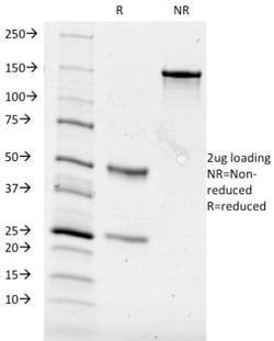
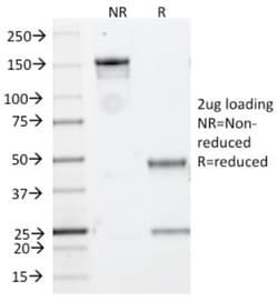
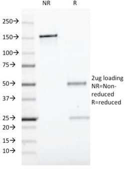
Molecular Weight of Antigen
44 kDa
Related Products


Description
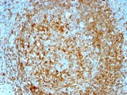
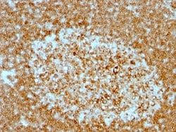
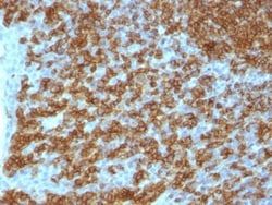
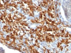
- Ensure accurate, reproducible results in Flow Cytometry, Immunohistochemistry (Paraffin), Immunofluorescence CD79A Monoclonal specifically detects CD79A in Human samples
- It is validated for Flow Cytometry, Immunohistochemistry, Immunocytochemistry/Immunofluorescence, Immunohistochemistry-Paraffin.
Compare Similar Items
Show Difference
Antigen: CD79A
Concentration: 0.2mg/mL
Applications: Flow Cytometry, Immunohistochemistry (Paraffin), Immunofluorescence
Conjugate: Unconjugated
Host Species: Mouse
Research Discipline: Adaptive Immunity, Cell Biology, Immunology
Formulation: 10mM PBS and 0.05% BSA with 0.05% Sodium Azide
Gene Alias: CD79a antigen, CD79A antigen (immunoglobulin-associated alpha), CD79a molecule, immunoglobulin-associated alpha, IGAB-cell antigen receptor complex-associated protein alpha chain, Ig-alpha, MB1, MB-1, MB-1 membrane glycoprotein, Membrane-bound immunoglobulin-associated protein, Surface IgM-associated protein
Gene Symbols: CD79A
Isotype: IgG1 κ
Purification Method: Protein A or G purified
Test Specificity: A disulphide-linked heterodimer, consisting of mb-1 (or CD79a) and B29 (or CD79b) polypeptides, is non-covalently associated with membrane-bound immunoglobulins on B cells. This complex of mb-1 and B29 polypeptides and immunoglobulin constitute the B cell Ag receptor. CD79a first appears at pre B cell stage, early in maturation, and persists until the plasma cell stage where it is found as an intracellular component. CD79a is found in the majority of acute leukemias of precursor B cell type, in B cell lines, B cell lymphomas, and in some myelomas. It is not present in myeloid or T cell lines. Anti-CD79a is generally used to complement anti-CD20 especially for mature B-cell lymphomas after treatment with Rituximab (anti-CD20). This antibody will stain many of the same lymphomas as anti-CD20, but also is more likely to stain B-lymphoblastic lymphoma/leukemia than is anti-CD20. Anti-CD79a also stains more cases of plasma cell myeloma and occasionally some types of endothelial cells as wel
Clone: IGA/515
Dilution: Flow Cytometry 0.5 - 1 ug/million cells in 0.1 ml, Immunohistochemistry-Paraffin 0.25 - 0.5 ug/ml, Immunofluorescence 0.5 - 1.0 ug/ml
Classification: Monoclonal
Form: Purified
Regulatory Status: RUO
Target Species: Human
Gene Accession No.: P11912
Gene ID (Entrez): 973
Immunogen: Recombinant full-length human IGA protein
Primary or Secondary: Primary
Content And Storage: Store at 4C.
Molecular Weight of Antigen: 44 kDa
Antigen: CD79A
Concentration: 0.2mg/mL
Applications: Flow Cytometry, Immunohistochemistry (Paraffin), Immunofluorescence
Conjugate: Unconjugated
Host Species: Mouse
Research Discipline: Adaptive Immunity, Cell Biology, Immunology
Formulation: 10mM PBS and 0.05% BSA with 0.05% Sodium Azide
Gene Alias: CD79a antigen, CD79A antigen (immunoglobulin-associated alpha), CD79a molecule, immunoglobulin-associated alpha, IGAB-cell antigen receptor complex-associated protein alpha chain, Ig-alpha, MB1, MB-1, MB-1 membrane glycoprotein, Membrane-bound immunoglobulin-associated protein, Surface IgM-associated protein
Gene Symbols: CD79A
Isotype: IgG1 κ
Purification Method: Protein A or G purified
Test Specificity: A disulphide-linked heterodimer, consisting of mb-1 (or CD79a) and B29 (or CD79b) polypeptides, is non-covalently associated with membrane-bound immunoglobulins on B cells. This complex of mb-1 and B29 polypeptides and immunoglobulin constitute the B cell Ag receptor. CD79a first appears at pre B cell stage, early in maturation, and persists until the plasma cell stage where it is found as an intracellular component. CD79a is found in the majority of acute leukemias of precursor B cell type, in B cell lines, B cell lymphomas, and in some myelomas. It is not present in myeloid or T cell lines. Anti-CD79a is generally used to complement anti-CD20 especially for mature B-cell lymphomas after treatment with Rituximab (anti-CD20). This antibody will stain many of the same lymphomas as anti-CD20, but also is more likely to stain B-lymphoblastic lymphoma/leukemia than is anti-CD20. Anti-CD79a also stains more cases of plasma cell myeloma and occasionally some types of endothelial cells as wel
Clone: IGA/764
Dilution: Flow Cytometry 0.5 - 1 ug/million cells in 0.1 ml, Immunohistochemistry-Paraffin 0.25 - 0.5 ug/ml, Immunofluorescence 0.5 - 1.0 ug/ml
Classification: Monoclonal
Form: Purified
Regulatory Status: RUO
Target Species: Human
Gene Accession No.: __
Gene ID (Entrez): 973
Immunogen: Recombinant human IGA protein
Primary or Secondary: Primary
Content And Storage: Store at 4C.
Molecular Weight of Antigen: 44 kDa

Antigen: CD84/SLAMF5
Concentration: 0.2 mg/mL
Applications: Flow Cytometry, SDS-Page, Immunofluorescence
Conjugate: Unconjugated
Host Species: Mouse
Research Discipline: __
Formulation: 10mM PBS and 0.05% BSA with 0.05% Sodium Azide
Gene Alias: CD84 antigen, CD84 antigen (leukocyte antigen), CD84 molecule, Cell surface antigen MAX.3, DKFZp781E2378, hCD84, hly9-beta, leucocyte differentiation antigen CD84, leukocyte antigen CD84, Leukocyte differentiation antigen CD84, mCD84, Signaling lymphocytic activation molecule 5, SLAM family member 5, SLAMF5LY9B
Gene Symbols: CD84
Isotype: IgG1 κ
Purification Method: Protein A or G purified
Test Specificity: Recognizes a protein of 74kDa, identified as CD84 (Workshop V; Code A085, C057). It is expressed on mature B cells and on B-cell lines, including pre-B-cell lines, but not on plasma cell lines. Immunohistochemical studies demonstrated that CD84 strongly expressed on tissues macrophages. CD84 is also highly expressed on platelets and, at low levels on peripheral blood T cells. It is a highly N-glycosylated protein and belongs to immunoglobulin superfamily. It may play a role in leukocyte activation.
Clone: 152-1D5
Dilution: Flow Cytometry 0.5 - 1 ug/million cells in 0.1 ml, SDS-Page, Immunofluorescence 0.5 - 1.0 ug/ml
Classification: Monoclonal
Form: Purified
Regulatory Status: RUO
Target Species: Human
Gene Accession No.: __
Gene ID (Entrez): 8832
Immunogen: Spleen cells of a patient with hairy cell leukemia
Primary or Secondary: Primary
Content And Storage: Store at 4C.
Molecular Weight of Antigen: 74 kDa