CD38 Antibody (FS02), Novus Biologicals™
Mouse Monoclonal Antibody
Manufacturer: Fischer Scientific
The price for this product is unavailable. Please request a quote
Antigen
CD38
Concentration
0.2 mg/mL
Applications
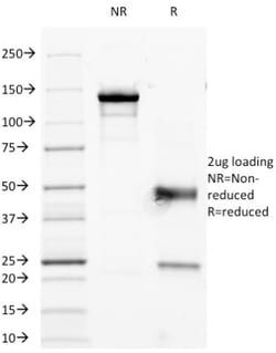
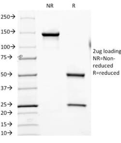
Flow Cytometry, SDS-Page, Immunofluorescence
Conjugate
Unconjugated
Host Species
Mouse
Research Discipline
Apoptosis, Cell Biology, Hematopoietic Stem Cell Markers, Immunology, Stem Cell Markers
Formulation
10mM PBS and 0.05% BSA with 0.05% Sodium Azide
Gene ID (Entrez)
952
Immunogen
Human CD38
Primary or Secondary
Primary
Content And Storage
Store at 4C.
Clone
FS02
Dilution
Flow Cytometry 0.5 - 1 ug/million cells in 0.1 ml, SDS-Page, Immunofluorescence 0.5 - 1.0 ug/ml
Classification
Monoclonal
Form
Purified
Regulatory Status
RUO
Target Species
Human
Gene Alias
ADP-ribosyl cyclase 1, ADP-ribosyl cyclase/cyclic ADP-ribose hydrolase, cADPr hydrolase 1, CD38 antigen, CD38 antigen (p45), CD38 molecule, Cyclic ADP-ribose hydrolase 1, EC 3.2.2.5, NAD(+) nucleosidase, T10
Gene Symbols
CD38
Isotype
IgG1 κ
Purification Method
Protein A or G purified
Test Specificity
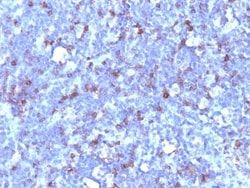
This antibody reacts with a type II membrane glycopeptide with a transmembrane sequence near the NH2-terminus. CD38 is a type II transmembrane glycoprotein that is present on early B- and T-cell lineages and activated B- and T-cells but is absent from most mature resting peripheral lymphocytes. CD38 is also found on thymocytes, pre-B cells, germinal center B-cells, mitogen-activated T-cells, monocytes and Ig-secreting plasma cells. CD38 is expressed on CD34+ cells. The CD34+CD38- population of hematopoietic stems cells defines the most pluripotent cells (e.g. blast colony forming cells).
Related Products

Description
- Ensure accurate, reproducible results in Flow Cytometry, Immunofluorescence CD38 Monoclonal specifically detects CD38 in Human samples
- It is validated for Flow Cytometry, Immunocytochemistry/Immunofluorescence, Immunofluorescence.
Compare Similar Items
Show Difference
Antigen: CD38
Concentration: 0.2 mg/mL
Applications: Flow Cytometry, SDS-Page, Immunofluorescence
Conjugate: Unconjugated
Host Species: Mouse
Research Discipline: Apoptosis, Cell Biology, Hematopoietic Stem Cell Markers, Immunology, Stem Cell Markers
Formulation: 10mM PBS and 0.05% BSA with 0.05% Sodium Azide
Gene ID (Entrez): 952
Immunogen: Human CD38
Primary or Secondary: Primary
Content And Storage: Store at 4C.
Clone: FS02
Dilution: Flow Cytometry 0.5 - 1 ug/million cells in 0.1 ml, SDS-Page, Immunofluorescence 0.5 - 1.0 ug/ml
Classification: Monoclonal
Form: Purified
Regulatory Status: RUO
Target Species: Human
Gene Alias: ADP-ribosyl cyclase 1, ADP-ribosyl cyclase/cyclic ADP-ribose hydrolase, cADPr hydrolase 1, CD38 antigen, CD38 antigen (p45), CD38 molecule, Cyclic ADP-ribose hydrolase 1, EC 3.2.2.5, NAD(+) nucleosidase, T10
Gene Symbols: CD38
Isotype: IgG1 κ
Purification Method: Protein A or G purified
Test Specificity: This antibody reacts with a type II membrane glycopeptide with a transmembrane sequence near the NH2-terminus. CD38 is a type II transmembrane glycoprotein that is present on early B- and T-cell lineages and activated B- and T-cells but is absent from most mature resting peripheral lymphocytes. CD38 is also found on thymocytes, pre-B cells, germinal center B-cells, mitogen-activated T-cells, monocytes and Ig-secreting plasma cells. CD38 is expressed on CD34+ cells. The CD34+CD38- population of hematopoietic stems cells defines the most pluripotent cells (e.g. blast colony forming cells).
Antigen: CD43/Sialophorin
Concentration: 0.2mg/mL
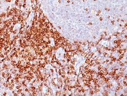
Applications: Flow Cytometry, Immunohistochemistry (Paraffin), Immunofluorescence
Conjugate: Unconjugated
Host Species: Mouse
Research Discipline: Immunology
Formulation: 10mM PBS and 0.05% BSA with 0.05% Sodium Azide
Gene ID (Entrez): 6693
Immunogen: Immature pluripotent human leukemia K562 cells
Primary or Secondary: Primary
Content And Storage: Store at 4C.
Clone: Bra7G
Dilution: Flow Cytometry 0.5 - 1 ug/million cells in 0.1 ml, Immunohistochemistry-Paraffin 0.5 - 1.0 ug/ml, Immunofluorescence 1 - 2 ug/ml
Classification: Monoclonal
Form: Purified
Regulatory Status: RUO
Target Species: Human
Gene Alias: CD43 antigen, CD43), Galactoglycoprotein, GALGP, Leukocyte sialoglycoprotein, Sialophorin, sialophorin (gpL115, leukosialin, CD43)
Gene Symbols: SPN
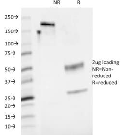
Isotype: IgM κ
Purification Method: PEG purified
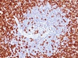
Test Specificity: It recognizes a cell surface glycoprotein of 95/115/135kDa (depending upon the extent of glycosylation), identified as CD43 (Workshop V). Epitope of MAb Bra7G is clearly different from that of MAb DF-T1, called b as opposed to a for DF-T1. 70-90% of T-cell lymphomas and from 22-37% of B-cell lymphomas express CD43. No reactivity has been observed with reactive B-cells. So a B-lineage population that co-expresses CD43 is highly likely to be a malignant lymphoma, especially a low-grade lymphoma, rather than a reactive B-cell population. When CD43 antibody is used in combination with anti-CD20, effective immunophenotyping of the lymphomas in formalin-fixed tissues can be obtained. Co-staining of a lymphoid infiltrate with anti-CD20 and anti-CD43 argues against a reactive process and favors a diagnosis of lymphoma.
Antigen: CD43/Sialophorin
Concentration: 0.2mg/mL
Applications: Flow Cytometry, Immunohistochemistry (Paraffin), Immunofluorescence
Conjugate: Unconjugated
Host Species: Mouse
Research Discipline: Immunology
Formulation: 10mM PBS and 0.05% BSA with 0.05% Sodium Azide
Gene ID (Entrez): 6693
Immunogen: Immature pluripotent human leukemia K562 cells
Primary or Secondary: Primary
Content And Storage: Store at 4C.
Clone: Bra7G
Dilution: Flow Cytometry 0.5 - 1 ug/million cells in 0.1 ml, Immunohistochemistry-Paraffin 0.5 - 1.0 ug/ml, Immunofluorescence 1 - 2 ug/ml
Classification: Monoclonal
Form: Purified
Regulatory Status: RUO
Target Species: Human
Gene Alias: CD43 antigen, CD43), Galactoglycoprotein, GALGP, Leukocyte sialoglycoprotein, Sialophorin, sialophorin (gpL115, leukosialin, CD43)
Gene Symbols: SPN
Isotype: IgM κ
Purification Method: PEG purified
Test Specificity: It recognizes a cell surface glycoprotein of 95/115/135kDa (depending upon the extent of glycosylation), identified as CD43 (Workshop V). Epitope of MAb Bra7G is clearly different from that of MAb DF-T1, called b as opposed to a for DF-T1. 70-90% of T-cell lymphomas and from 22-37% of B-cell lymphomas express CD43. No reactivity has been observed with reactive B-cells. So a B-lineage population that co-expresses CD43 is highly likely to be a malignant lymphoma, especially a low-grade lymphoma, rather than a reactive B-cell population. When CD43 antibody is used in combination with anti-CD20, effective immunophenotyping of the lymphomas in formalin-fixed tissues can be obtained. Co-staining of a lymphoid infiltrate with anti-CD20 and anti-CD43 argues against a reactive process and favors a diagnosis of lymphoma.