CD48/SLAMF2 Antibody (5-4.8), Novus Biologicals™
Mouse Monoclonal Antibody
Manufacturer: Fischer Scientific
The price for this product is unavailable. Please request a quote
Antigen
CD48/SLAMF2
Concentration
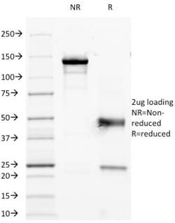
0.2 mg/mL
Applications
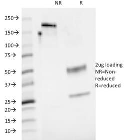
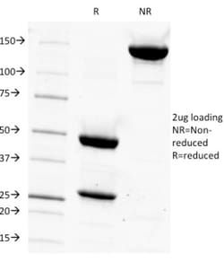
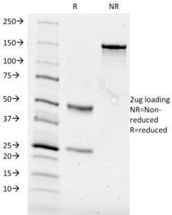
Flow Cytometry, SDS-Page, Immunofluorescence
Conjugate
Unconjugated
Host Species
Mouse
Research Discipline
Cell Biology, Cellular Markers, Immunology, Stem Cell Markers
Formulation
10mM PBS and 0.05% BSA with 0.05% Sodium Azide
Gene ID (Entrez)
962
Immunogen
Human peripheral blood lymphocytes
Primary or Secondary
Primary
Content And Storage
Store at 4C.
Molecular Weight of Antigen
45 kDa
Clone
5-4.8
Dilution
Flow Cytometry 0.5 - 1 ug/million cells in 0.1 ml, SDS-Page, Immunofluorescence 0.5 - 1.0 ug/ml
Classification
Monoclonal
Form
Purified
Regulatory Status
RUO
Target Species
Human
Gene Alias
BCM1 surface antigen, BCM1Leukocyte antigen MEM-102, BLAST, BLAST1TCT.1, B-lymphocyte activation marker BLAST-1, CD48 antigen, CD48 antigen (B-cell membrane protein), CD48 molecule, hCD48, mCD48, MEM-102, SLAMF2
Gene Symbols
CD48
Isotype
IgG2a κ
Purification Method
Protein A or G purified
Test Specificity
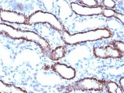
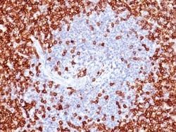
Reacts with human CD48, a 45kDa glycosyl phophatidyl-inositol (GPI)-anchored cell surface protein. CD48 is strongly expressed on lymphocytes and monocytes and weakly on granulocytes but is absent on platelets, fibroblasts, epithelium and endothelium. CD48 is one of the markers for detecting the defects of GPI anchoring structure on the patients with paroxysmal nocturnal hemoglobulinuria (PNH) and serves as a low affinity ligand for CD2.
Related Products

Description
- Ensure accurate, reproducible results in Flow Cytometry, Immunofluorescence CD48/SLAMF2 Monoclonal specifically detects CD48/SLAMF2 in Human samples
- It is validated for Flow Cytometry, Immunocytochemistry/Immunofluorescence, Immunofluorescence.
Compare Similar Items
Show Difference
Antigen: CD48/SLAMF2
Concentration: 0.2 mg/mL
Applications: Flow Cytometry, SDS-Page, Immunofluorescence
Conjugate: Unconjugated
Host Species: Mouse
Research Discipline: Cell Biology, Cellular Markers, Immunology, Stem Cell Markers
Formulation: 10mM PBS and 0.05% BSA with 0.05% Sodium Azide
Gene ID (Entrez): 962
Immunogen: Human peripheral blood lymphocytes
Primary or Secondary: Primary
Content And Storage: Store at 4C.
Molecular Weight of Antigen: 45 kDa
Clone: 5-4.8
Dilution: Flow Cytometry 0.5 - 1 ug/million cells in 0.1 ml, SDS-Page, Immunofluorescence 0.5 - 1.0 ug/ml
Classification: Monoclonal
Form: Purified
Regulatory Status: RUO
Target Species: Human
Gene Alias: BCM1 surface antigen, BCM1Leukocyte antigen MEM-102, BLAST, BLAST1TCT.1, B-lymphocyte activation marker BLAST-1, CD48 antigen, CD48 antigen (B-cell membrane protein), CD48 molecule, hCD48, mCD48, MEM-102, SLAMF2
Gene Symbols: CD48
Isotype: IgG2a κ
Purification Method: Protein A or G purified
Test Specificity: Reacts with human CD48, a 45kDa glycosyl phophatidyl-inositol (GPI)-anchored cell surface protein. CD48 is strongly expressed on lymphocytes and monocytes and weakly on granulocytes but is absent on platelets, fibroblasts, epithelium and endothelium. CD48 is one of the markers for detecting the defects of GPI anchoring structure on the patients with paroxysmal nocturnal hemoglobulinuria (PNH) and serves as a low affinity ligand for CD2.
Antigen: CA19-9/Sialyl Lewisa
Concentration: 0.2mg/mL
Applications: Flow Cytometry, Immunohistochemistry (Paraffin), Immunofluorescence
Conjugate: Unconjugated
Host Species: Mouse
Research Discipline: __
Formulation: 10mM PBS and 0.05% BSA with 0.05% Sodium Azide
Gene ID (Entrez): __
Immunogen: Precipitin lines obtained after immuno-diffusion using MAb 116-NS-19-9 and mucins isolated from an ovarian cyst of a Lewis A+B- patient (0Le).
Primary or Secondary: Primary
Content And Storage: Store at 4C.
Molecular Weight of Antigen: __
Clone: 121SLE
Dilution: Flow Cytometry 0.5 - 1ug/million cells, Immunohistochemistry-Paraffin 1:50-1:100, Immunofluorescence 1:25 - 1:50
Classification: Monoclonal
Form: Purified
Regulatory Status: RUO
Target Species: Human
Gene Alias: __
Gene Symbols: __
Isotype: IgM κ
Purification Method: Protein A or G purified
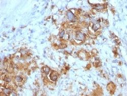
Test Specificity: CA19-9, a carbohydrate epitope expressed on a high MW (>400kDa) mucin glycoprotein, is a sialyl Lewisa structure which is synthesized from type 1 blood group precursor chains and is present in individuals expressing the Lewisa and/or Lewisb blood group antigens. In normal tissues, sialyl Lewisa antigen is present in ductal epithelium of the breast, kidney, salivary gland, and sweat glands. Its expression is greatly enhanced in serum as well as in the majority of tumor cells in gastrointestinal (GI) carcinomas, including adenocarcinomas of the stomach, intestine, and pancreas. Preoperative elevated CA19-9 levels in patients with stage I pancreatic carcinoma decrease to normal values following surgery. When used serially, CA19-9 can predict recurrence of disease prior to radiographic or clinical findings. This MAb is excellent for staining of formalin-fixed, paraffin-embedded tissues.
Antigen: CA19-9/Sialyl Lewisa
Concentration: 0.2mg/mL
Applications: Flow Cytometry, Immunohistochemistry (Paraffin), Immunofluorescence
Conjugate: Unconjugated
Host Species: Mouse
Research Discipline: __
Formulation: 10mM PBS and 0.05% BSA with 0.05% Sodium Azide
Gene ID (Entrez): __
Immunogen: Precipitin lines obtained after immuno-diffusion using MAb 116-NS-19-9 and mucins isolated from an ovarian cyst of a Lewis A+B- patient (0Le).
Primary or Secondary: Primary
Content And Storage: Store at 4C.
Molecular Weight of Antigen: __
Clone: 121SLE
Dilution: Flow Cytometry 0.5 - 1ug/million cells, Immunohistochemistry-Paraffin 1:50-1:100, Immunofluorescence 1:25 - 1:50
Classification: Monoclonal
Form: Purified
Regulatory Status: RUO
Target Species: Human
Gene Alias: __
Gene Symbols: __
Isotype: IgM κ
Purification Method: Protein A or G purified
Test Specificity: CA19-9, a carbohydrate epitope expressed on a high MW (>400kDa) mucin glycoprotein, is a sialyl Lewisa structure which is synthesized from type 1 blood group precursor chains and is present in individuals expressing the Lewisa and/or Lewisb blood group antigens. In normal tissues, sialyl Lewisa antigen is present in ductal epithelium of the breast, kidney, salivary gland, and sweat glands. Its expression is greatly enhanced in serum as well as in the majority of tumor cells in gastrointestinal (GI) carcinomas, including adenocarcinomas of the stomach, intestine, and pancreas. Preoperative elevated CA19-9 levels in patients with stage I pancreatic carcinoma decrease to normal values following surgery. When used serially, CA19-9 can predict recurrence of disease prior to radiographic or clinical findings. This MAb is excellent for staining of formalin-fixed, paraffin-embedded tissues.