Cadherin-16 Antibody (CDH16/1071), Novus Biologicals™
Mouse Monoclonal Antibody has been used in 2 publications
Manufacturer: Fischer Scientific
The price for this product is unavailable. Please request a quote
Antigen
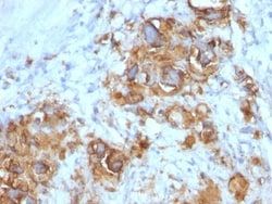
Cadherin-16
Concentration
0.2mg/mL
Applications
Flow Cytometry, Immunohistochemistry (Paraffin), Immunofluorescence
Conjugate
Unconjugated
Host Species
Mouse
Target Species
Human, Mouse, Rat, Canine, Rabbit
Gene Accession No.
O75309
Gene ID (Entrez)
1014
Immunogen
Recombinant human CDH16 protein
Primary or Secondary
Primary
Content And Storage
Store at 4C.
Molecular Weight of Antigen
130 kDa
Clone
CDH16/1071
Dilution
Flow Cytometry 0.5 - 1 ug/million cells in 0.1 ml, Immunohistochemistry-Paraffin 0.5 - 1.0 ug/ml, Immunofluorescence 1 - 2 ug/ml
Classification
Monoclonal
Form
Purified
Regulatory Status
RUO
Formulation
10mM PBS and 0.05% BSA with 0.05% Sodium Azide
Gene Alias
cadherin 16, KSP-cadherin, cadherin-16, Kidney-specific cadherin, KSP-cadherin
Gene Symbols
CDH16
Isotype
IgG1 κ
Purification Method
Protein A or G purified
Test Specificity
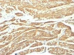
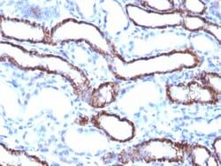
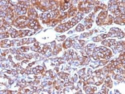
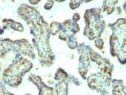
This MAb recognizes a protein of 130kDa, identified as Ksp-cadherin. Cadherins form a superfamily of related glycoproteins that mediate calcium-dependent cell adhesion and transmit signals from the extracellular matrix to the cytoplasm. Cadherins have been implicated in embryogenesis, tissue morphogenesis, tissue structure maintenance, cell polarization, neoplastic invasiveness and metastasis, and membrane transport. It is suggested that Ksp-cadherin is a marker for terminal differentiation of the basolateral membranes of renal tubular epithelial cells. Within the kidney, Ksp-Cadherin is found exclusively in the basolateral membrane of renal tubular epithelial cells and collecting duct cells, and not in glomeruli, renal interstitial cells, or blood vessels.Ksp-Cadherin has been suggested to distinguish Chromophobe Renal-Cell Carcinoma from Oncocytoma.
Related Products

Description
- Ensure accurate, reproducible results in Flow Cytometry, Immunohistochemistry (Paraffin), Immunofluorescence Cadherin-16 Monoclonal specifically detects Cadherin-16 in Human, Mouse, Rat, Canine, Rabbit samples
- It is validated for Western Blot, Flow Cytometry, ELISA, Immunohistochemistry, Immunohistochemistry-Paraffin.
Compare Similar Items
Show Difference
Antigen: Cadherin-16
Concentration: 0.2mg/mL
Applications: Flow Cytometry, Immunohistochemistry (Paraffin), Immunofluorescence
Conjugate: Unconjugated
Host Species: Mouse
Target Species: Human, Mouse, Rat, Canine, Rabbit
Gene Accession No.: O75309
Gene ID (Entrez): 1014
Immunogen: Recombinant human CDH16 protein
Primary or Secondary: Primary
Content And Storage: Store at 4C.
Molecular Weight of Antigen: 130 kDa
Clone: CDH16/1071
Dilution: Flow Cytometry 0.5 - 1 ug/million cells in 0.1 ml, Immunohistochemistry-Paraffin 0.5 - 1.0 ug/ml, Immunofluorescence 1 - 2 ug/ml
Classification: Monoclonal
Form: Purified
Regulatory Status: RUO
Formulation: 10mM PBS and 0.05% BSA with 0.05% Sodium Azide
Gene Alias: cadherin 16, KSP-cadherin, cadherin-16, Kidney-specific cadherin, KSP-cadherin
Gene Symbols: CDH16
Isotype: IgG1 κ
Purification Method: Protein A or G purified
Test Specificity: This MAb recognizes a protein of 130kDa, identified as Ksp-cadherin. Cadherins form a superfamily of related glycoproteins that mediate calcium-dependent cell adhesion and transmit signals from the extracellular matrix to the cytoplasm. Cadherins have been implicated in embryogenesis, tissue morphogenesis, tissue structure maintenance, cell polarization, neoplastic invasiveness and metastasis, and membrane transport. It is suggested that Ksp-cadherin is a marker for terminal differentiation of the basolateral membranes of renal tubular epithelial cells. Within the kidney, Ksp-Cadherin is found exclusively in the basolateral membrane of renal tubular epithelial cells and collecting duct cells, and not in glomeruli, renal interstitial cells, or blood vessels.Ksp-Cadherin has been suggested to distinguish Chromophobe Renal-Cell Carcinoma from Oncocytoma.
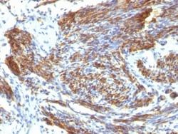
Antigen: Caldesmon/CALD1
Concentration: 0.2mg/mL
Applications: Flow Cytometry, Immunohistochemistry (Paraffin), Immunofluorescence
Conjugate: Unconjugated
Host Species: Mouse
Target Species: Human, Rat (Negative)
Gene Accession No.: Q05682
Gene ID (Entrez): 800
Immunogen: Recombinant full-length human CALD1 protein
Primary or Secondary: Primary
Content And Storage: Store at 4C.
Molecular Weight of Antigen: 150 kDa
Clone: CALD1/820
Dilution: Flow Cytometry 0.5 - 1 ug/million cells in 0.1 ml, Immunohistochemistry-Paraffin 0.25 - 0.5 ug/ml, Immunofluorescence 1 - 2 ug/ml
Classification: Monoclonal
Form: Purified
Regulatory Status: RUO
Formulation: 10mM PBS and 0.05% BSA with 0.05% Sodium Azide
Gene Alias: CAD, caldesmon, caldesmon 1, CDMH-CAD, HCAD, LCAD, L-CAD, MGC21352, NAG22
Gene Symbols: CALD1
Isotype: IgG1 κ
Purification Method: Protein A or G purified
Test Specificity: Recognizes a protein of 150kDa, which is identified as the high molecular weight variant of Caldesmon. Two closely related variants of human caldesmon have been identified which are different in their electrophoretic mobility and cellular distribution. The h-caldesmon variant (120-150kDa) is predominantly expressed in smooth muscle whereas l-caldesmon (70-80kDa) is found in non- muscle tissue and cells. Neither of the two variants has been detected in skeletal muscle. This MAb recognizes only the 150kDa variant (h-caldesmon) in Western blots of human aortic media extracts and is unreactive with fibroblast extracts from cultivated human foreskin. Caldesmon is a developmentally regulated protein involved in smooth muscle and non-muscle contraction.