Moesin Antibody (MSN/492), Novus Biologicals™
Manufacturer: Fischer Scientific
Select a Size
| Pack Size | SKU | Availability | Price |
|---|---|---|---|
| Each of 1 | NBP24457900-Each-of-1 | In Stock | ₹ 23,852.00 |
NBP24457900 - Each of 1
In Stock
Quantity
1
Base Price: ₹ 23,852.00
GST (18%): ₹ 4,293.36
Total Price: ₹ 28,145.36
Antigen
Moesin
Classification
Monoclonal
Concentration
0.2mg/mL
Dilution
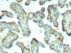
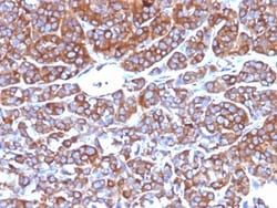
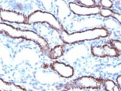
Western Blot 0.5 - 1.0 ug/ml, Flow Cytometry 0.5 - 1 ug/million cells in 0.1 ml, Immunohistochemistry-Paraffin 0.5 - 1.0 ug/ml, Immunofluorescence 0.5 - 1.0 ug/ml
Gene Accession No.
P26038
Gene Symbols
MSN
Immunogen
Recombinant full-length human Moesin protein
Purification Method
Protein A or G purified
Regulatory Status
RUO
Primary or Secondary
Primary
Test Specificity
Recognizes 78kDa moesin protein. Moesin, a member of the talin-4.1 superfamily, is a linking protein of the sub-membranous actin cytoskeleton. It is expressed in variable amounts in cells of different phenotypes such as macrophages, lymphocytes, fibroblastic, endothelial, epithelial, and neuronal cell lines but not in blood cells. The ERM proteins, ezrin, radixin, and moesin are involved in a variety of cellular functions, such as cell adhesion, migration, and the organization of cell surface structures, and are highly homologous, both in protein sequence and in functional activity, with merlin/schwannomin, a neurofibromatosis-2-associated tumor-suppressor protein. Cell lines of epithelial and mesothelial origin contain both moesin and radixin whereas cells of endothelial and lymphoid origin express moesin.
Content And Storage
Store at 4C.
Isotype
IgG1 κ
Applications
Western Blot, Flow Cytometry, Immunohistochemistry (Paraffin), Immunofluorescence
Clone
MSN/492
Conjugate
Unconjugated
Formulation
10mM PBS and 0.05% BSA with 0.05% Sodium Azide
Gene Alias
Membrane-organizing extension spike protein, moesin
Host Species
Mouse
Molecular Weight of Antigen
78 kDa
Quantity
0.02 mg
Research Discipline
Cytoskeleton Markers, Stem Cell Markers
Gene ID (Entrez)
4478
Target Species
Human, Rat (Negative)
Form
Purified
Related Products

Description
- Ensure accurate, reproducible results in Western Blot, Flow Cytometry, Immunohistochemistry (Paraffin), Immunofluorescence Moesin Monoclonal specifically detects Moesin in Human, Rat (Negative) samples
- It is validated for Western Blot, Flow Cytometry, Immunohistochemistry, Immunocytochemistry/Immunofluorescence, Immunohistochemistry-Paraffin, Knockout Validated.
Compare Similar Items
Show Difference
Antigen: Moesin
Classification: Monoclonal
Concentration: 0.2mg/mL
Dilution: Western Blot 0.5 - 1.0 ug/ml, Flow Cytometry 0.5 - 1 ug/million cells in 0.1 ml, Immunohistochemistry-Paraffin 0.5 - 1.0 ug/ml, Immunofluorescence 0.5 - 1.0 ug/ml
Gene Accession No.: P26038
Gene Symbols: MSN
Immunogen: Recombinant full-length human Moesin protein
Purification Method: Protein A or G purified
Regulatory Status: RUO
Primary or Secondary: Primary
Test Specificity: Recognizes 78kDa moesin protein. Moesin, a member of the talin-4.1 superfamily, is a linking protein of the sub-membranous actin cytoskeleton. It is expressed in variable amounts in cells of different phenotypes such as macrophages, lymphocytes, fibroblastic, endothelial, epithelial, and neuronal cell lines but not in blood cells. The ERM proteins, ezrin, radixin, and moesin are involved in a variety of cellular functions, such as cell adhesion, migration, and the organization of cell surface structures, and are highly homologous, both in protein sequence and in functional activity, with merlin/schwannomin, a neurofibromatosis-2-associated tumor-suppressor protein. Cell lines of epithelial and mesothelial origin contain both moesin and radixin whereas cells of endothelial and lymphoid origin express moesin.
Content And Storage: Store at 4C.
Isotype: IgG1 κ
Applications: Western Blot, Flow Cytometry, Immunohistochemistry (Paraffin), Immunofluorescence
Clone: MSN/492
Conjugate: Unconjugated
Formulation: 10mM PBS and 0.05% BSA with 0.05% Sodium Azide
Gene Alias: Membrane-organizing extension spike protein, moesin
Host Species: Mouse
Molecular Weight of Antigen: 78 kDa
Quantity: 0.02 mg
Research Discipline: Cytoskeleton Markers, Stem Cell Markers
Gene ID (Entrez): 4478
Target Species: Human, Rat (Negative)
Form: Purified
Antigen: Moesin
Classification: Monoclonal
Concentration: 0.2mg/mL
Dilution: Western Blot 0.5 - 1.0 ug/ml, Flow Cytometry 0.5 - 1 ug/million cells in 0.1 ml, Immunohistochemistry-Paraffin 0.5 - 1.0 ug/ml, Immunofluorescence 0.5 - 1.0 ug/ml
Gene Accession No.: P26038
Gene Symbols: MSN
Immunogen: Recombinant full-length human Moesin protein
Purification Method: Protein A or G purified
Regulatory Status: RUO
Primary or Secondary: Primary
Test Specificity: Recognizes 78kDa moesin protein. Moesin, a member of the talin-4.1 superfamily, is a linking protein of the sub-membranous actin cytoskeleton. It is expressed in variable amounts in cells of different phenotypes such as macrophages, lymphocytes, fibroblastic, endothelial, epithelial, and neuronal cell lines but not in blood cells. The ERM proteins, ezrin, radixin, and moesin are involved in a variety of cellular functions, such as cell adhesion, migration, and the organization of cell surface structures, and are highly homologous, both in protein sequence and in functional activity, with merlin/schwannomin, a neurofibromatosis-2-associated tumor-suppressor protein. Cell lines of epithelial and mesothelial origin contain both moesin and radixin whereas cells of endothelial and lymphoid origin express moesin.
Content And Storage: Store at 4C.
Isotype: IgG1 κ
Applications: Western Blot, Flow Cytometry, Immunohistochemistry (Paraffin), Immunofluorescence
Clone: MSN/492
Conjugate: Unconjugated
Formulation: 10mM PBS and 0.05% BSA with 0.05% Sodium Azide
Gene Alias: Membrane-organizing extension spike protein, moesin
Host Species: Mouse
Molecular Weight of Antigen: 78 kDa
Quantity: 0.1 mg
Research Discipline: Cytoskeleton Markers, Stem Cell Markers
Gene ID (Entrez): 4478
Target Species: Human, Rat (Negative)
Form: Purified
Antigen: Moesin
Classification: Monoclonal
Concentration: 0.2mg/mL
Dilution: Western Blot 0.5 - 1.0 ug/ml, Flow Cytometry 0.5 - 1 ug/million cells in 0.1 ml, Immunohistochemistry-Paraffin 0.5 - 1.0 ug/ml, Immunofluorescence 0.5 - 1.0 ug/ml
Gene Accession No.: P26038
Gene Symbols: MSN
Immunogen: Recombinant full-length human Moesin protein
Purification Method: Protein A or G purified
Regulatory Status: RUO
Primary or Secondary: Primary
Test Specificity: Recognizes 78kDa moesin protein. Moesin, a member of the talin-4.1 superfamily, is a linking protein of the sub-membranous actin cytoskeleton. It is expressed in variable amounts in cells of different phenotypes such as macrophages, lymphocytes, fibroblastic, endothelial, epithelial, and neuronal cell lines but not in blood cells. The ERM proteins, ezrin, radixin, and moesin are involved in a variety of cellular functions, such as cell adhesion, migration, and the organization of cell surface structures, and are highly homologous, both in protein sequence and in functional activity, with merlin/schwannomin, a neurofibromatosis-2-associated tumor-suppressor protein. Cell lines of epithelial and mesothelial origin contain both moesin and radixin whereas cells of endothelial and lymphoid origin express moesin.
Content And Storage: Store at 4C.
Isotype: IgG1 κ
Applications: Western Blot, Flow Cytometry, Immunohistochemistry (Paraffin), Immunofluorescence
Clone: MSN/492
Conjugate: Unconjugated
Formulation: 10mM PBS and 0.05% BSA with 0.05% Sodium Azide
Gene Alias: Membrane-organizing extension spike protein, moesin
Host Species: Mouse
Molecular Weight of Antigen: 78 kDa
Quantity: 0.2 mg
Research Discipline: Cytoskeleton Markers, Stem Cell Markers
Gene ID (Entrez): 4478
Target Species: Human, Rat (Negative)
Form: Purified