CD14 Antibody (LPSR/927), Novus Biologicals™
Mouse Monoclonal Antibody
Manufacturer: Fischer Scientific
The price for this product is unavailable. Please request a quote
Antigen
CD14
Concentration
0.2 mg/mL
Applications
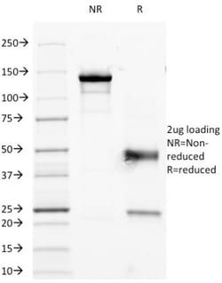
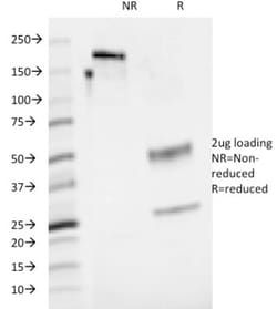
Flow Cytometry, SDS-Page, Immunofluorescence
Conjugate
Unconjugated
Host Species
Mouse
Research Discipline
Adaptive Immunity, Apoptosis, Asthma, Biologically Active Proteins, Cancer, Cell Biology, Cytokine Research, Immunology, Innate Immunity, Myeloid Cell Markers, Stem Cell Markers
Formulation
10mM PBS and 0.05% BSA with 0.05% Sodium Azide
Gene ID (Entrez)
929
Immunogen
Recombinant human CD14 protein
Primary or Secondary
Primary
Content And Storage
Store at 4C.
Molecular Weight of Antigen
55 kDa
Clone
LPSR/927
Dilution
Flow Cytometry 0.5 - 1 ug/million cells in 0.1 ml, SDS-Page, Immunofluorescence 0.5 - 1.0 ug/ml
Classification
Monoclonal
Form
Purified
Regulatory Status
RUO
Target Species
Human
Gene Alias
CD14 antigen, CD14 molecule, monocyte differentiation antigen CD14, Myeloid cell-specific leucine-rich glycoprotein
Gene Symbols
CD14
Isotype
IgG1 κ
Purification Method
Protein A or G purified
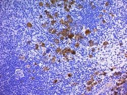
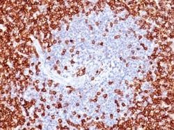
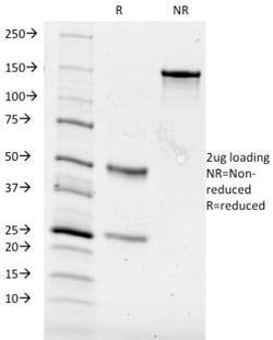
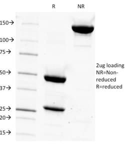
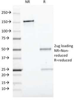
Test Specificity
Recognizes a protein of 55kDa, identified as CD14 (also known lipopolysaccharide receptor). CD14 is expressed strongly on monocytes and macrophage and weakly on the surface of neutrophils. CD14 is anchored to cells by linkage to glycosylphosphatidylinositol (GPI) and functions as a high affinity receptor for complexes of LPS and LPS binding protein (LBP). Soluble CD14, also binding to LPS, acts at physiological concentration as an LPS agonist and has, at higher concentrations, an LPS antagonizing effect in cell activation.
Related Products

Description
- Ensure accurate, reproducible results in Flow Cytometry, Immunofluorescence CD14 Monoclonal specifically detects CD14 in Human samples
- It is validated for Flow Cytometry, Immunocytochemistry/Immunofluorescence, Immunofluorescence.
Compare Similar Items
Show Difference
Antigen: CD14
Concentration: 0.2 mg/mL
Applications: Flow Cytometry, SDS-Page, Immunofluorescence
Conjugate: Unconjugated
Host Species: Mouse
Research Discipline: Adaptive Immunity, Apoptosis, Asthma, Biologically Active Proteins, Cancer, Cell Biology, Cytokine Research, Immunology, Innate Immunity, Myeloid Cell Markers, Stem Cell Markers
Formulation: 10mM PBS and 0.05% BSA with 0.05% Sodium Azide
Gene ID (Entrez): 929
Immunogen: Recombinant human CD14 protein
Primary or Secondary: Primary
Content And Storage: Store at 4C.
Molecular Weight of Antigen: 55 kDa
Clone: LPSR/927
Dilution: Flow Cytometry 0.5 - 1 ug/million cells in 0.1 ml, SDS-Page, Immunofluorescence 0.5 - 1.0 ug/ml
Classification: Monoclonal
Form: Purified
Regulatory Status: RUO
Target Species: Human
Gene Alias: CD14 antigen, CD14 molecule, monocyte differentiation antigen CD14, Myeloid cell-specific leucine-rich glycoprotein
Gene Symbols: CD14
Isotype: IgG1 κ
Purification Method: Protein A or G purified
Test Specificity: Recognizes a protein of 55kDa, identified as CD14 (also known lipopolysaccharide receptor). CD14 is expressed strongly on monocytes and macrophage and weakly on the surface of neutrophils. CD14 is anchored to cells by linkage to glycosylphosphatidylinositol (GPI) and functions as a high affinity receptor for complexes of LPS and LPS binding protein (LBP). Soluble CD14, also binding to LPS, acts at physiological concentration as an LPS agonist and has, at higher concentrations, an LPS antagonizing effect in cell activation.
Antigen: CD19
Concentration: 0.2 mg/mL
Applications: Flow Cytometry, SDS-Page, Immunofluorescence
Conjugate: Unconjugated
Host Species: Mouse
Research Discipline: Adaptive Immunity, Cytokine Research, Immunology, Innate Immunity, Mesenchymal Stem Cell Markers, Signal Transduction, Stem Cell Lines, Stem Cell Markers
Formulation: 10mM PBS and 0.05% BSA with 0.05% Sodium Azide
Gene ID (Entrez): 930
Immunogen: Recombinant human CD19 protein
Primary or Secondary: Primary
Content And Storage: Store at 4C.
Molecular Weight of Antigen: 95 kDa
Clone: CVID3/429
Dilution: Flow Cytometry 0.5 - 1 ug/million cells in 0.1 ml, SDS-Page, Immunofluorescence 0.5 - 1.0 ug/ml
Classification: Monoclonal
Form: Purified
Regulatory Status: RUO
Target Species: Human, Monkey, Primate
Gene Alias: B4, B-lymphocyte antigen CD19, B-lymphocyte surface antigen B4, CD19 antigen, CD19 molecule, CVID3, Differentiation antigen CD19, MGC12802, T-cell surface antigen Leu-12
Gene Symbols: CD19
Isotype: IgG1 κ
Purification Method: Protein A or G purified
Test Specificity: CD19 is a transmembrane glycoprotein that contains two extracellular immunoglobulin-like domains. CD19 is present in both benign and malignant B-cells and is considered to be the most reliable surface marker of this lineage over a wide range of maturational stages. In normal lymphoid tissue, CD19 is observed in germinal centers, in mantle zone cells, and in scattered cells of the inter-follicular areas. Anti-CD19 exhibits an overall immunoreactivity pattern similar to those of the antibodies against CD20 and CD22. However, in contrast to CD20, expression of CD19 is continuous throughout B-cell development and through terminal differentiation of B-cells into plasma cells. Anti-CD19 positivity is seen in the vast majority of B-cell neoplasms commonly at a lower intensity than normal B-cell counterparts. Plasma cell neoplasms are nearly always negative, as are T-cell neoplasms
Antigen: CD27 Ligand/TNFSF7/CD70
Concentration: 0.2 mg/mL
Applications: Flow Cytometry, Immunoprecipitation, Immunohistochemistry (Frozen), Immunofluorescence
Conjugate: Unconjugated
Host Species: Mouse
Research Discipline: Apoptosis
Formulation: 10mM PBS and 0.05% BSA with 0.05% Sodium Azide
Gene ID (Entrez): 970
Immunogen: Human WM-1 (Waldenstrom s macroglobulinemia) cell line
Primary or Secondary: Primary
Content And Storage: Store at 4C.
Molecular Weight of Antigen: 30 kDa
Clone: BU69
Dilution: Flow Cytometry 0.5 - 1 ug/million cells in 0.1 ml, Immunoprecipitation 0.5 - 1 ug/500ug Protein Lysate, Immunohistochemistry-Frozen 0.5 - 1 ug/ml, Immunofluorescence 0.5 - 1.0 ug/ml
Classification: Monoclonal
Form: Purified
Regulatory Status: RUO
Target Species: Human
Gene Alias: CD27-L, CD27LCD27 ligand, CD27LG, CD70, CD70 molecule, Ki-24 antigen, surface antigen CD70, TNFSF7CD70 antigen, tumor necrosis factor (ligand) superfamily, member 7, Tumor necrosis factor ligand superfamily member 7
Gene Symbols: CD70
Isotype: IgG1 κ
Purification Method: Protein A or G purified
Test Specificity: It recognizes a protein of 30kDa, identified as CD70. It is a cytokine that belongs to the tumor necrosis factor (TNF) ligand family. This cytokine is a ligand for TNFRSF27/CD27. It is a surface antigen on activated, but not on resting, T- and B-lymphocytes. It induces proliferation of co-stimulated T cells, enhances the generation of cytolytic T cells, and contributes to T cell activation. This cytokine is also reported to play a role in regulating B-cell activation, cytotoxic function of natural killer cells, and immunoglobulin synthesis. This MAb blocks the interaction between CD27 and CD70, and has been shown to inhibit T cell proliferation induced by dendritic cells.