CD7 Antibody (T3-3A1), Novus Biologicals™
Mouse Monoclonal Antibody
Manufacturer: Fischer Scientific
The price for this product is unavailable. Please request a quote
Antigen
CD7
Concentration
0.2 mg/mL
Applications
Flow Cytometry, Immunofluorescence
Conjugate
Unconjugated
Host Species
Mouse
Research Discipline
Cytokine Research, Signal Transduction
Formulation
10mM PBS and 0.05% BSA with 0.05% Sodium Azide
Gene ID (Entrez)
924
Immunogen
Human T cells
Primary or Secondary
Primary
Content And Storage
Store at 4C.
Molecular Weight of Antigen
40 kDa
Clone
T3-3A1
Dilution
Flow Cytometry 0.5 - 1 ug/million cells in 0.1 ml, Immunofluorescence 0.5 - 1.0 ug/ml
Classification
Monoclonal
Form
Purified
Regulatory Status
RUO
Target Species
Human
Gene Alias
CD7 antigen, CD7 antigen (p41), CD7 molecule, GP40T-cell surface antigen Leu-9, LEU-9, T-cell antigen CD7, T-cell leukemia antigen, Tp40, TP41p41 protein
Gene Symbols
CD7
Isotype
IgG1 κ
Purification Method
Protein A or G purified
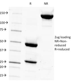
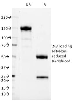
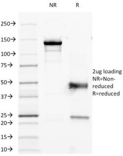
Test Specificity
Recognizes a protein of 40kDa, identified as CD7, a member of the immunoglobulin gene superfamily. Its N-terminal amino acids 1-107 are highly homologous to Ig kappa-L chains whereas the carboxyl-terminal region of the extracellular domain is proline-rich and has been postulated to form a stalk from which the Ig domain projects. CD7 is expressed on the majority of immature and mature T-lymphocytes, and T cell leukemia. It is also found on natural killer cells, a small subpopulation of normal B cells and on malignant B cells. Cross-linking surface CD7 positively modulates T cell and NK cell activity as measured by calcium fluxes, expression of adhesion molecules, cytokine secretion and proliferation. CD7 associates directly with phosphoinositol 3'-kinase. CD7 ligation induces production of D-3 phosphoinositides and tyrosine phosphorylation.
Related Products

Description
- Ensure accurate, reproducible results in Flow Cytometry, Immunofluorescence CD7 Monoclonal specifically detects CD7 in Human samples
- It is validated for Flow Cytometry, Immunocytochemistry/Immunofluorescence, Immunofluorescence.
Compare Similar Items
Show Difference
Antigen: CD7
Concentration: 0.2 mg/mL
Applications: Flow Cytometry, Immunofluorescence
Conjugate: Unconjugated
Host Species: Mouse
Research Discipline: Cytokine Research, Signal Transduction
Formulation: 10mM PBS and 0.05% BSA with 0.05% Sodium Azide
Gene ID (Entrez): 924
Immunogen: Human T cells
Primary or Secondary: Primary
Content And Storage: Store at 4C.
Molecular Weight of Antigen: 40 kDa
Clone: T3-3A1
Dilution: Flow Cytometry 0.5 - 1 ug/million cells in 0.1 ml, Immunofluorescence 0.5 - 1.0 ug/ml
Classification: Monoclonal
Form: Purified
Regulatory Status: RUO
Target Species: Human
Gene Alias: CD7 antigen, CD7 antigen (p41), CD7 molecule, GP40T-cell surface antigen Leu-9, LEU-9, T-cell antigen CD7, T-cell leukemia antigen, Tp40, TP41p41 protein
Gene Symbols: CD7
Isotype: IgG1 κ
Purification Method: Protein A or G purified
Test Specificity: Recognizes a protein of 40kDa, identified as CD7, a member of the immunoglobulin gene superfamily. Its N-terminal amino acids 1-107 are highly homologous to Ig kappa-L chains whereas the carboxyl-terminal region of the extracellular domain is proline-rich and has been postulated to form a stalk from which the Ig domain projects. CD7 is expressed on the majority of immature and mature T-lymphocytes, and T cell leukemia. It is also found on natural killer cells, a small subpopulation of normal B cells and on malignant B cells. Cross-linking surface CD7 positively modulates T cell and NK cell activity as measured by calcium fluxes, expression of adhesion molecules, cytokine secretion and proliferation. CD7 associates directly with phosphoinositol 3'-kinase. CD7 ligation induces production of D-3 phosphoinositides and tyrosine phosphorylation.
Antigen: CD8 beta
Concentration: 0.2 mg/mL
Applications: Flow Cytometry, Immunofluorescence
Conjugate: Unconjugated
Host Species: Mouse
Research Discipline: Adaptive Immunity, Cytokine Research, Immunology, Innate Immunity, Signal Transduction, Stem Cell Markers
Formulation: 10mM PBS and 0.05% BSA with 0.05% Sodium Azide
Gene ID (Entrez): 926
Immunogen: Human CD8 beta
Primary or Secondary: Primary
Content And Storage: Store at 4C.
Molecular Weight of Antigen: 32 kDa
Clone: BU88
Dilution: Flow Cytometry 0.5 - 1 ug/million cells in 0.1 ml, Immunofluorescence 0.5 - 1.0 ug/ml
Classification: Monoclonal
Form: Purified
Regulatory Status: RUO
Target Species: Human
Gene Alias: CD8 antigen, beta polypeptide 1 (p37), CD8b antigen, CD8b molecule, CD8B1P37, LEU2, LY3, LYT3, MGC119115, T lymphocyte surface glycoprotein beta chain, T-cell surface glycoprotein CD8 beta chain
Gene Symbols: CD8B
Isotype: IgG1 κ
Purification Method: Protein A or G purified
Test Specificity: The T cell receptor (TCR) is a heterodimer composed of either alpha and beta or gamma and delta chains. CD3 chains and the CD4 or CD8 co-receptors are also required for efficient signal transduction through the TCR. The TCR is expressed on T helper and T cytotoxic cells that can be distinguished by their expression of CD4 and CD8. T helper cells express CD4 proteins and T cytotoxic cells display CD8. CD8 (also designated Leu 2 or T8), a cell surface glycoprotein, is a two chain complex (alpha-alpha or alpha-beta) receptor that binds class I MHC molecules presented by the antigen-presenting cell (APC). A primary function of CD8 is to facilitate antigen recognition by the TCR and to strengthen the avidity of the TCR-antigen interactions. An additional role for CD8-expressing T cells may be to maintain low levels of HIV expression.
Antigen: CD8 alpha
Concentration: 0.2 mg/mL
Applications: Flow Cytometry, Immunofluorescence
Conjugate: Unconjugated
Host Species: Mouse
Research Discipline: Innate Immunity
Formulation: 10mM PBS and 0.05% BSA with 0.05% Sodium Azide
Gene ID (Entrez): 925
Immunogen: Human peripheral lymphocytes
Primary or Secondary: Primary
Content And Storage: Store at 4C.
Molecular Weight of Antigen: 32 kDa
Clone: RIV11
Dilution: Flow Cytometry 0.5 - 1 ug/million cells in 0.1 ml, Immunofluorescence 0.5 - 1.0 ug/ml
Classification: Monoclonal
Form: Purified
Regulatory Status: RUO
Target Species: Human
Gene Alias: CD8, CD8 antigen, alpha polypeptide (p32), CD8a antigen, CD8a molecule, Leu2, Leu2 T-lymphocyte antigen, MAL, OKT8 T-cell antigen, p32, RPA-T8, RPA-T8 antibody flow, RPA-T8 CD8, RPA-T8 Clone, RPA-T8 Flow, T cell co-receptor, T8 T-cell antigen, T-cell antigen Leu2, T-cell surface glycoprotein CD8 alpha chain, T-lymphocyte differentiation antigen T8/Leu-2
Gene Symbols: CD8A
Isotype: IgG1 κ
Purification Method: Protein A or G purified
Test Specificity: Recognizes a protein of 32kDa, identified as CD8a (also known as CD8 chain, T cell co-receptor, Leu2, and T8). CD8 molecule consists of two chains, termed and chain, which are expressed as a disulphide-linked heterodimer or as an homodimer. CD8 is expressed on T cell subset (cytotoxic/suppressor T cells), thymocytes and NK cells. The majority of CD8+ T-cells expresses CD8 as heterodimer. Some subpopulation of CD8+ T cells as well as NK cells may express homodimer. CD8 functions as a co-receptor in concert with TCR for binding the MHC class I/peptide complex. The HIV-2 envelope glycoprotein binds CD8 chain (but not chain). The cytoplasmic domain of CD8 associates with p56lck tyrosine kinase.