CD90/Thy1 Antibody (AF-9), Novus Biologicals™
Mouse Monoclonal Antibody
Manufacturer: Fischer Scientific
The price for this product is unavailable. Please request a quote
Antigen
CD90/Thy1
Concentration
0.2 mg/mL
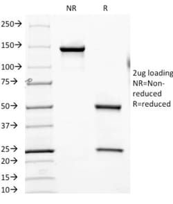
Applications
Flow Cytometry, SDS-Page, Immunofluorescence
Conjugate
Unconjugated
Host Species
Mouse
Research Discipline
Hematopoietic Stem Cell Markers, Hepatic Stem Cell Markers, Immunology, Innate Immunity, Mesenchymal Stem Cell Markers, Signal Transduction, Stem Cell Markers
Formulation
10mM PBS and 0.05% BSA with 0.05% Sodium Azide
Gene Alias
CD90, CD90 antigen, CDw90, FLJ33325, Thy-1 antigen, Thy-1 cell surface antigen, thy-1 membrane glycoprotein, Thy-1 T-cell antigen
Gene Symbols
THY1
Isotype
IgG1 κ
Purification Method
Protein A or G purified
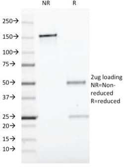
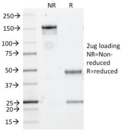
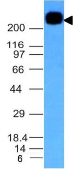
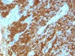
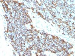
Test Specificity
Recognizes a protein of 18-35kDa, identified as CD90 (also known as Thy1). CD90 is a member of the immunoglobulin superfamily. It may contribute to inhibition of proliferation/differentiation of hematopoietic stem cells and neuron memory formation in the CNS. It consists of a single Ig domain (112 amino acids; 25-35 kDa) inserted into the cell membrane via a GPI anchor. Expressed by hematopoietic stem cells and neurons in all species studied. Its highly expressed in connective tissue and various fibroblast and stromal cell lines, expressed on all thymocytes and peripheral T cells in mice, but in humans expressed only on small % fetal thymocytes, 10-40% of CD34+ cells in bone marrow, and <1% of CD3+CD4+ lymphocytes in peripheral circulation. It is also expressed by human lymph node HEV endothelium but not other endothelia. Lastly, it is expressed by a limited number of lymphoblastoid and leukemic cell lines.
Clone
AF-9
Dilution
Flow Cytometry 0.5 - 1 ug/million cells in 0.1 ml, SDS-Page, Immunofluorescence 0.5 - 1.0 ug/ml
Classification
Monoclonal
Form
Purified
Regulatory Status
RUO
Target Species
Human, Bovine
Gene Accession No.
P04216
Gene ID (Entrez)
7070
Immunogen
Human T-lymphoma cells
Primary or Secondary
Primary
Content And Storage
Store at 4C.
Related Products

Description
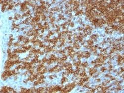
- Ensure accurate, reproducible results in Flow Cytometry, Immunofluorescence CD90/Thy1 Monoclonal specifically detects CD90/Thy1 in Human, Bovine samples
- It is validated for Flow Cytometry, Immunocytochemistry/Immunofluorescence, Immunofluorescence.
Compare Similar Items
Show Difference
Antigen: CD90/Thy1
Concentration: 0.2 mg/mL
Applications: Flow Cytometry, SDS-Page, Immunofluorescence
Conjugate: Unconjugated
Host Species: Mouse
Research Discipline: Hematopoietic Stem Cell Markers, Hepatic Stem Cell Markers, Immunology, Innate Immunity, Mesenchymal Stem Cell Markers, Signal Transduction, Stem Cell Markers
Formulation: 10mM PBS and 0.05% BSA with 0.05% Sodium Azide
Gene Alias: CD90, CD90 antigen, CDw90, FLJ33325, Thy-1 antigen, Thy-1 cell surface antigen, thy-1 membrane glycoprotein, Thy-1 T-cell antigen
Gene Symbols: THY1
Isotype: IgG1 κ
Purification Method: Protein A or G purified
Test Specificity: Recognizes a protein of 18-35kDa, identified as CD90 (also known as Thy1). CD90 is a member of the immunoglobulin superfamily. It may contribute to inhibition of proliferation/differentiation of hematopoietic stem cells and neuron memory formation in the CNS. It consists of a single Ig domain (112 amino acids; 25-35 kDa) inserted into the cell membrane via a GPI anchor. Expressed by hematopoietic stem cells and neurons in all species studied. Its highly expressed in connective tissue and various fibroblast and stromal cell lines, expressed on all thymocytes and peripheral T cells in mice, but in humans expressed only on small % fetal thymocytes, 10-40% of CD34+ cells in bone marrow, and <1% of CD3+CD4+ lymphocytes in peripheral circulation. It is also expressed by human lymph node HEV endothelium but not other endothelia. Lastly, it is expressed by a limited number of lymphoblastoid and leukemic cell lines.
Clone: AF-9
Dilution: Flow Cytometry 0.5 - 1 ug/million cells in 0.1 ml, SDS-Page, Immunofluorescence 0.5 - 1.0 ug/ml
Classification: Monoclonal
Form: Purified
Regulatory Status: RUO
Target Species: Human, Bovine
Gene Accession No.: P04216
Gene ID (Entrez): 7070
Immunogen: Human T-lymphoma cells
Primary or Secondary: Primary
Content And Storage: Store at 4C.
Antigen: CD99
Concentration: 0.2mg/mL
Applications: Flow Cytometry, Immunohistochemistry (Paraffin), Immunofluorescence
Conjugate: Unconjugated
Host Species: Mouse
Research Discipline: Immunology
Formulation: No buffer with 0.05% Sodium Azide
Gene Alias: 12E7, antigen identified by monoclonal antibodies 12E7, F21 and O13, CD99 antigenY homolog, CD99 molecule, E2 antigen, HBA71, MIC2 (monoclonal 12E7), MIC2Y, MSK5X, Protein MIC2, surface antigen MIC2, T-cell surface glycoprotein E2
Gene Symbols: CD99
Isotype: IgG
Purification Method: Tissue culture supernatant
Test Specificity: Recognizes a sialoglycoprotein of 27-32kDa, identified as CD99, or MIC2 gene product, or E2 antigen. MIC2 gene is located in the pseudo-autosomal region of the human X and Y chromosome. MIC2 gene encodes two distinct proteins, which are produced by alternative splicing of the CD99 gene transcript and are identified as bands of 30 and 32kDa (p30/32). Although its function is not fully understood, CD99 is implicated in various cellular processes including homotypic aggregation of T cells, upregulation of T cell receptor and MHS molecules, apoptosis of immature thymocytes and leukocyte diapedesis.CD99 is expressed on the cell membrane of some lymphocytes, cortical thymocytes, and granulosa cells of the ovary. Most pancreatic islet cells, Sertoli cells of the testis, and some endothelial cells express this antigen. Mature granulocytes express very little or no CD99. MIC2 is strongly expressed on Ewings sarcoma cells and primitive peripheral neuroectodermal tumors.
Clone: 12E7 + MIC2/877
Dilution: Flow Cytometry 5 - 10 ul/million cells in 0.1ml, Immunohistochemistry-Paraffin 1:100-1:200, Immunofluorescence 1:100-1:200
Classification: Monoclonal
Form: Supernatant
Regulatory Status: RUO
Target Species: Human
Gene Accession No.: P14209, P14209
Gene ID (Entrez): 4267
Immunogen: Human acute lymphocytic leukemia T-cells (12E7); Recombinant human MIC2 protein (MIC2/877)
Primary or Secondary: Primary
Content And Storage: Store at 4C.
Antigen: CD6
Concentration: 0.2 mg/mL
Applications: Flow Cytometry, SDS-Page, Immunofluorescence
Conjugate: Unconjugated
Host Species: Mouse
Research Discipline: Immunology
Formulation: 10mM PBS and 0.05% BSA with 0.05% Sodium Azide
Gene Alias: CD6 antigenFLJ44171, CD6 molecule, T12, Tp120, TP120T-cell differentiation antigen CD6
Gene Symbols: CD6
Isotype: IgG1
Purification Method: Protein A or G purified
Test Specificity: CD6 is a type I transmembrane glycoprotein that contains a 24-amino acid signal sequence, three extracellular scavenger receptor cysteine-rich (SRCR) domains, a membrane-spanning domain and a 44-amino acid cytoplasmic domain. The CD6 glycoprotein is tyrosine phosphorylated during TCR-mediated T cell activation. CD6 shows significant homology to CD5. CD6 is present on mature thymocytes, peripheral T cells and a subset of B cells. Antibodies to CD6 are used to deplete T cells from bone marrow transplants to prevent graft versus host disease.
Clone: SPV-L14
Dilution: Flow Cytometry 0.5 - 1 ug/million cells in 0.1 ml, SDS-Page, Immunofluorescence 0.5 - 1.0 ug/ml
Classification: Monoclonal
Form: Purified
Regulatory Status: RUO
Target Species: Human, Mouse
Gene Accession No.: __
Gene ID (Entrez): 923
Immunogen: CD8+ cytotoxic T-cell clone
Primary or Secondary: Primary
Content And Storage: Store at 4C.