PTH Antibody (SPM604) - N-terminal, Novus Biologicals™
Mouse Monoclonal Antibody
Manufacturer: Fischer Scientific
The price for this product is unavailable. Please request a quote
Antigen
PTH
Concentration
0.2mg/mL
Applications
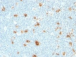
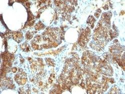
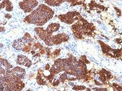
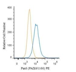
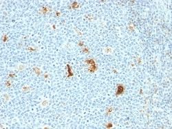
Flow Cytometry, Immunohistochemistry (Paraffin), Immunofluorescence
Conjugate
Unconjugated
Host Species
Mouse
Research Discipline
Apoptosis, Cancer
Formulation
10mM PBS and 0.05% BSA with 0.05% Sodium Azide
Gene ID (Entrez)
5741
Immunogen
A synthetic peptide from the N-terminal of human PTH polypeptide.
Primary or Secondary
Primary
Content And Storage
Store at 4C.
Molecular Weight of Antigen
9 kDa
Clone
SPM604
Dilution
Flow Cytometry 0.5 - 1 ug/million cells in 0.1 ml, Immunohistochemistry-Paraffin 0.5 - 1.0 ug/ml, Immunofluorescence 0.5 - 1.0 ug/ml
Classification
Monoclonal
Form
Purified
Regulatory Status
RUO
Target Species
Human
Gene Alias
Parathormone, Parathyrin, parathyroid hormone, parathyroid hormone 1, PTH1
Gene Symbols
PTH
Isotype
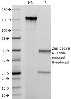
IgG2b κ
Purification Method
Protein A or G purified
Test Specificity
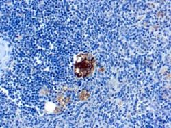
Epitope of this MAb maps in the N-terminus of PTH, a hormone produced by the parathyroid gland that regulates the concentration of calcium and phosphorus in extracellular fluid. This hormone elevates blood Ca2+ levels by dissolving the salts in bone and preventing their renal excretion.It is produced in the parathyroid gland as an 84 amino acid single chain polypeptide. It can also be secreted as N-terminal truncated fragments or C-terminal fragments after intracellular degradation, as in case of hypercalcemia. Defects in this gene are a cause of familial isolated hypoparathyroidism (FIH); also called autosomal dominant hypoparathyroidism or autosomal dominant hypocalcemia. FIH is characterized by hypocalcemia and hyperphosphatemia due to inadequate secretion of parathyroid hormone. Symptoms are seizures, tetany and cramps. FIH exist both as autosomal dominant and recessive forms of hypoparathyroidism.
Related Products
Description
- Ensure accurate, reproducible results in Flow Cytometry, Immunohistochemistry (Paraffin), Immunofluorescence PTH Monoclonal specifically detects PTH in Human samples
- It is validated for Immunohistochemistry, Immunohistochemistry-Paraffin.
Compare Similar Items
Show Difference
Antigen: PTH
Concentration: 0.2mg/mL
Applications: Flow Cytometry, Immunohistochemistry (Paraffin), Immunofluorescence
Conjugate: Unconjugated
Host Species: Mouse
Research Discipline: Apoptosis, Cancer
Formulation: 10mM PBS and 0.05% BSA with 0.05% Sodium Azide
Gene ID (Entrez): 5741
Immunogen: A synthetic peptide from the N-terminal of human PTH polypeptide.
Primary or Secondary: Primary
Content And Storage: Store at 4C.
Molecular Weight of Antigen: 9 kDa
Clone: SPM604
Dilution: Flow Cytometry 0.5 - 1 ug/million cells in 0.1 ml, Immunohistochemistry-Paraffin 0.5 - 1.0 ug/ml, Immunofluorescence 0.5 - 1.0 ug/ml
Classification: Monoclonal
Form: Purified
Regulatory Status: RUO
Target Species: Human
Gene Alias: Parathormone, Parathyrin, parathyroid hormone, parathyroid hormone 1, PTH1
Gene Symbols: PTH
Isotype: IgG2b κ
Purification Method: Protein A or G purified
Test Specificity: Epitope of this MAb maps in the N-terminus of PTH, a hormone produced by the parathyroid gland that regulates the concentration of calcium and phosphorus in extracellular fluid. This hormone elevates blood Ca2+ levels by dissolving the salts in bone and preventing their renal excretion.It is produced in the parathyroid gland as an 84 amino acid single chain polypeptide. It can also be secreted as N-terminal truncated fragments or C-terminal fragments after intracellular degradation, as in case of hypercalcemia. Defects in this gene are a cause of familial isolated hypoparathyroidism (FIH); also called autosomal dominant hypoparathyroidism or autosomal dominant hypocalcemia. FIH is characterized by hypocalcemia and hyperphosphatemia due to inadequate secretion of parathyroid hormone. Symptoms are seizures, tetany and cramps. FIH exist both as autosomal dominant and recessive forms of hypoparathyroidism.
Antigen: S100A8/A9
Concentration: 0.2 mg/mL
Applications: Flow Cytometry, Immunohistochemistry (Paraffin), SDS-Page, Immunofluorescence
Conjugate: Unconjugated
Host Species: Mouse
Research Discipline: Cancer
Formulation: 1.0mM PBS and 0.05% BSA with 0.05% Sodium Azide
Gene ID (Entrez): 6279
Immunogen: Affinity purified monocyte membrane preparation
Primary or Secondary: Primary
Content And Storage: Store at 4C.
Molecular Weight of Antigen: __
Clone: MAC387
Dilution: Flow Cytometry 0.5 - 1 ug/million cells in 0.1 ml, Immunohistochemistry-Paraffin 0.5 - 1.0 ug/ml, SDS-Page, Immunofluorescence 0.5 - 1.0 ug/ml
Classification: Monoclonal
Form: Purified
Regulatory Status: RUO
Target Species: Human, Mouse, Rat, Porcine, Baboon, Canine, Equine, Feline, Guinea Pig, Goat, Monkey, Rabbit
Gene Alias: 60B8AG, CAGA, CFAG, CGLA, CP-10, L1Ag, MA387, MIF, MRP8, NIF, P8, S100 calcium binding protein A8, S100A8
Gene Symbols: S100A8
Isotype: IgG1 κ
Purification Method: Protein A or G purified
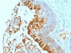
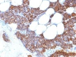
Test Specificity: Recognizes the L1 or Calprotectin molecule, an intra-cytoplasmic antigen comprising of a 12kDa alpha chain and a 14kDa beta chain expressed by granulocytes, monocytes and by tissue macrophages. Macrophages usually arise from hematopoietic stem cells in the bone marrow. Under migration into tissues, the monocytes undergo further differentiation to become multifunctional tissue macrophages. They are classified into normal and inflammatory macrophages. Normal macrophages include macrophages in connective tissue (histiocytes), liver (Kupffer's cells), lung (alveolar macrophages), lymph nodes (free and fixed macrophages), spleen (free and fixed macrophages), bone marrow (fixed macrophages), serous fluids (pleural and peritoneal macrophages), skin (histiocytes, Langerhans's cell) and in other tissues. Inflammatory macrophages are present in various exudates. Macrophages are part of the innate immune system, recognizing, engulfing and destroying many potential pathogens including bacteria, pa
Antigen: S100A8/A9
Concentration: 0.2 mg/mL
Applications: Flow Cytometry, Immunohistochemistry (Paraffin), Immunofluorescence
Conjugate: Unconjugated
Host Species: Mouse
Research Discipline: Cancer
Formulation: 1.0mM PBS and 0.05% BSA with 0.05% Sodium Azide
Gene ID (Entrez): 6279
Immunogen: Affinity purified monocyte membrane preparation
Primary or Secondary: Primary
Content And Storage: Store at 4C.
Molecular Weight of Antigen: __
Clone: SPM281
Dilution: Flow Cytometry 0.5 - 1 ug/million cells in 0.1 ml, Immunohistochemistry-Paraffin 0.5 - 1.0 ug/ml, Immunofluorescence 0.5 - 1.0 ug/ml
Classification: Monoclonal
Form: Purified
Regulatory Status: RUO
Target Species: Human, Mouse, Rat, Porcine, Baboon, Canine, Equine, Feline, Guinea Pig, Goat, Monkey, Rabbit
Gene Alias: 60B8AG, CAGA, CFAG, CGLA, CP-10, L1Ag, MA387, MIF, MRP8, NIF, P8, S100 calcium binding protein A8, S100A8
Gene Symbols: S100A8
Isotype: IgG1 κ
Purification Method: Protein A or G purified
Test Specificity: Recognizes the L1 or Calprotectin molecule, an intra-cytoplasmic antigen comprising of a 12kDa alpha chain and a 14kDa beta chain expressed by granulocytes, monocytes and by tissue macrophages. Macrophages usually arise from hematopoietic stem cells in the bone marrow. Under migration into tissues, the monocytes undergo further differentiation to become multifunctional tissue macrophages. They are classified into normal and inflammatory macrophages. Normal macrophages include macrophages in connective tissue (histiocytes), liver (Kupffer s cells), lung (alveolar macrophages), lymph nodes (free and fixed macrophages), spleen (free and fixed macrophages), bone marrow (fixed macrophages), serous fluids (pleural and peritoneal macrophages), skin (histiocytes, Langerhans's cell) and in other tissues. Inflammatory macrophages are present in various exudates. Macrophages are part of the innate immune system, recognizing, engulfing and destroying many potential pathogens including bacteria, pa