G-CSF Antibody (SPM468), Novus Biologicals™
Mouse Monoclonal Antibody
Manufacturer: Fischer Scientific
The price for this product is unavailable. Please request a quote
Antigen
G-CSF
Concentration
0.2mg/mL
Applications
Flow Cytometry, Immunohistochemistry (Paraffin), Immunofluorescence
Conjugate
Unconjugated
Host Species
Mouse
Research Discipline
Cell Cycle and Replication, Cytokine Research
Formulation
10mM PBS and 0.05% BSA with 0.05% Sodium Azide
Gene Alias
C17orf33, chromosome 17 open reading frame 33, colony stimulating factor 3 (granulocyte), CSF3OS, filgrastim, G-CSF, GCSFlenograstim, granulocyte colony-stimulating factor, Lenograstim, MGC45931, pluripoietin
Gene Symbols
CSF3
Isotype
IgG1
Purification Method
Protein A or G purified
Test Specificity
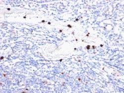
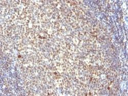
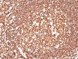
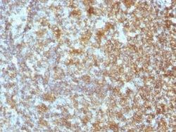
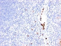
This MAb recognizes granulocyte-colony stimulating factor (G-CSF) in the cytoplasm of mature granulocytes. It shows no reactivity with any other cell types. Markers of myeloid cells are useful in the identification of different levels of cellular differentiation. It reacts with early precursor and mature forms of myeloid cells. It is useful for the detection of myeloid leukemias and granulocytic sarcomas. It can be used as a marker of granulocytes in normal tissues or inflammatory processes.G-CSF is a pleiotropic cytokine that influences differentiation, proliferation and activation of the neutrophilic granulocyte lineage. The human G-CSF cDNA encodes a 207 amino acid precursor containing a 29 amino acid signal peptide that is proteolytically cleaved to form a 178 amino acid residue mature protein. Two G-CSF s, which are identical except for a three amino acid deletion in the amino-terminus of one form of the protein have been isolated from human cells. Murine and human G-CSF s share 7
Clone
SPM468
Dilution
Flow Cytometry 0.5 - 1 ug/million cells in 0.1 ml, Immunohistochemistry-Paraffin 0.5 - 1.0 ug/ml, Immunofluorescence 1 - 2 ug/ml
Classification
Monoclonal
Form
Purified
Regulatory Status
RUO
Target Species
Human, Monkey
Gene Accession No.
P09919
Gene ID (Entrez)
1440
Immunogen
Nuclei from pokeweed mitogen stimulated human peripheral blood lymphocytes
Primary or Secondary
Primary
Content And Storage
Store at 4C.
Molecular Weight of Antigen
19 kDa
Related Products


Description
- Ensure accurate, reproducible results in Flow Cytometry, Immunohistochemistry (Paraffin), Immunofluorescence G-CSF Monoclonal specifically detects G-CSF in Human, Monkey, Rhesus Macaque samples
- It is validated for Flow Cytometry, Immunohistochemistry, Immunocytochemistry/Immunofluorescence, Immunohistochemistry-Paraffin, Immunofluorescence.
Compare Similar Items
Show Difference
Antigen: G-CSF
Concentration: 0.2mg/mL
Applications: Flow Cytometry, Immunohistochemistry (Paraffin), Immunofluorescence
Conjugate: Unconjugated
Host Species: Mouse
Research Discipline: Cell Cycle and Replication, Cytokine Research
Formulation: 10mM PBS and 0.05% BSA with 0.05% Sodium Azide
Gene Alias: C17orf33, chromosome 17 open reading frame 33, colony stimulating factor 3 (granulocyte), CSF3OS, filgrastim, G-CSF, GCSFlenograstim, granulocyte colony-stimulating factor, Lenograstim, MGC45931, pluripoietin
Gene Symbols: CSF3
Isotype: IgG1
Purification Method: Protein A or G purified
Test Specificity: This MAb recognizes granulocyte-colony stimulating factor (G-CSF) in the cytoplasm of mature granulocytes. It shows no reactivity with any other cell types. Markers of myeloid cells are useful in the identification of different levels of cellular differentiation. It reacts with early precursor and mature forms of myeloid cells. It is useful for the detection of myeloid leukemias and granulocytic sarcomas. It can be used as a marker of granulocytes in normal tissues or inflammatory processes.G-CSF is a pleiotropic cytokine that influences differentiation, proliferation and activation of the neutrophilic granulocyte lineage. The human G-CSF cDNA encodes a 207 amino acid precursor containing a 29 amino acid signal peptide that is proteolytically cleaved to form a 178 amino acid residue mature protein. Two G-CSF s, which are identical except for a three amino acid deletion in the amino-terminus of one form of the protein have been isolated from human cells. Murine and human G-CSF s share 7
Clone: SPM468
Dilution: Flow Cytometry 0.5 - 1 ug/million cells in 0.1 ml, Immunohistochemistry-Paraffin 0.5 - 1.0 ug/ml, Immunofluorescence 1 - 2 ug/ml
Classification: Monoclonal
Form: Purified
Regulatory Status: RUO
Target Species: Human, Monkey
Gene Accession No.: P09919
Gene ID (Entrez): 1440
Immunogen: Nuclei from pokeweed mitogen stimulated human peripheral blood lymphocytes
Primary or Secondary: Primary
Content And Storage: Store at 4C.
Molecular Weight of Antigen: 19 kDa
Antigen: Glycophorin A
Concentration: 0.2mg/mL
Applications: Western Blot, Flow Cytometry, Immunohistochemistry (Paraffin), Immunofluorescence
Conjugate: Unconjugated
Host Species: Mouse
Research Discipline: Cancer, Signal Transduction
Formulation: 10mM PBS and 0.05% BSA with 0.05% Sodium Azide
Gene Alias: CD235a antigen, glycophorin A (includes MN blood group), glycophorin A (MNS blood group), glycophorin Erik, glycophorin MiI, glycophorin MiIII, glycophorin MiV, glycophorin MiX, glycophorin SAT, glycophorin Sta type C, glycophorin-A, GPA, GPErik, GpMiIII, GPSAT, HGpMiIII, HGpMiV, HGpMiX, HGpMiXI, HGpSta(C), Mi.V glycoprotein (24 AA), MN sialoglycoprotein, MNS, PAS-2, recombinant glycophorin A-B Miltenberger-DR
Gene Symbols: GYPA
Isotype: IgG1 κ
Purification Method: Protein A or G purified
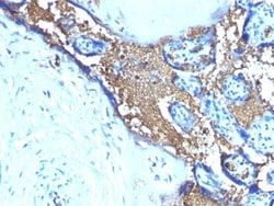
Test Specificity: Recognizes a sialoglycoprotein of 39kDa, identified as glycophorin A (GPA). It is present on red blood cells (RBC) and erythroid precursor cells. It has been shown that glycophorin acts as the receptor for Sandei virus and parvovirus. Glycophorins A (GPA) and B (GPB), which are single, trans-membrane sialoglycoproteins. GPA is the carrier of blood group M and N specificities, while GPB accounts for S and U specificities. GPA and GPB provide the cells with a large mucin like surface and it has been suggested this provides a barrier to cell fusion, so minimizing aggregation between red blood cells in the circulation.
Clone: SPM599
Dilution: Western Blot, Flow Cytometry 0.5 - 1 ug/million cells in 0.1 ml, Immunohistochemistry-Paraffin 0.25 - 0.5 ug/ml, Immunofluorescence 0.5 - 1.0 ug/ml
Classification: Monoclonal
Form: Purified
Regulatory Status: RUO
Target Species: Human
Gene Accession No.: P02724
Gene ID (Entrez): 2993
Immunogen: Recombinant human glycophorin A protein
Primary or Secondary: Primary
Content And Storage: Store at 4C.
Molecular Weight of Antigen: 39 kDa
Antigen: Glypican 3
Concentration: 0.2mg/mL
Applications: Flow Cytometry, Immunohistochemistry (Paraffin), Immunofluorescence
Conjugate: Unconjugated
Host Species: Mouse
Research Discipline: __
Formulation: 1.0mM PBS and 0.05% BSA with 0.05% Sodium Azide
Gene Alias: DGSX, glypican 3, glypican proteoglycan 3, glypican-3, GTR2-2, heparan sulphate proteoglycan, Intestinal protein OCI-5, MXR7, OCI5, OCI-5, secreted glypican-3, SGB, SGBS, SGBS1SDYS
Gene Symbols: GPC3
Isotype: IgG
Purification Method: Protein A or G purified
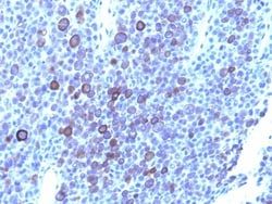
Test Specificity: Glypican-3 (GPC3) is an integral membrane protein that is mutated in the Simpson-Golabi-Behmel syndrome (SGBS). SGBS is characterized by pre- and post-natal overgrowth and is a recessive X-linked condition. GPC3 may also be found in a secreted form. Anti-GPC3 has been identified as a useful tumor marker for the diagnosis of hepatocellular carcinoma (HCC), hepatoblastoma, melanoma, testicular germ cell tumors, and Wilm s tumor. In patients with HCC, GPC3 is overexpressed in neoplastic liver tissue and elevated in serum, but is undetectable in normal liver, benign liver, and the serum of healthy donors. GPC3 expression is also found to be higher in HCC liver tissue than in cirrhotic liver or liver with focal lesions such as dysplastic nodules and areas of hepatic adenoma (HA) with malignant transformation. In the context of testicular germ cell tumors, GPC3 expression is up regulated in certain histologic subtypes, specifically yolk sac tumors and choriocarcinoma. A high level of GPC3 ex
Clone: 1G12 + GPC3/863
Dilution: Flow Cytometry 0.5 - 1 ug/million cells in 0.1 ml, Immunohistochemistry-Paraffin 0.5 - 1.0 ug/ml, Immunofluorescence 0.5 - 1.0 ug/ml
Classification: Monoclonal
Form: Purified
Regulatory Status: RUO
Target Species: Human
Gene Accession No.: P51654, P51654
Gene ID (Entrez): 2719
Immunogen: Recombinant fragment containing amino acids 511-580 of human glypican-3 (1G12); Recombinant full-length human GPC3 protein (GPC3/863)
Primary or Secondary: Primary
Content And Storage: Store at 4C.
Molecular Weight of Antigen: 67 kDa