NuMA Antibody (SPM300), Novus Biologicals™
Mouse Monoclonal Antibody
Manufacturer: Fischer Scientific
The price for this product is unavailable. Please request a quote
Antigen
NuMA
Concentration
0.2 mg/mL
Applications
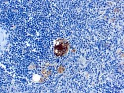
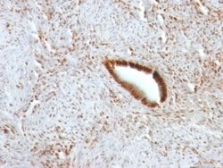
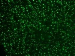
Flow Cytometry, Immunohistochemistry (Paraffin), Immunofluorescence
Conjugate
Unconjugated
Host Species
Mouse
Research Discipline
Breast Cancer, Cell Biology, Cell Cycle and Replication, Cellular Markers, Mitotic Regulators
Formulation
10mM PBS and 0.05% BSA with 0.05% Sodium Azide
Gene ID (Entrez)
4926
Immunogen
Colon carcinoma 174T cells
Primary or Secondary
Primary
Content And Storage
Store at 4C.
Molecular Weight of Antigen
228 kDa
Clone
SPM300
Dilution
Flow Cytometry 0.5 - 1 ug/million cells in 0.1 ml, Immunohistochemistry-Paraffin 0.5 - 1.0 ug/ml, Immunofluorescence 0.5 - 1.0 ug/ml
Classification
Monoclonal
Form
Purified
Regulatory Status
RUO
Target Species
Human
Gene Alias
centrophilin stabilizes mitotic spindle in mitotic cells, nuclear mitotic apparatus protein 1, NUMA, NuMA protein, SP-H antigen, structural nuclear protein
Gene Symbols
NUMA1
Isotype
IgM κ
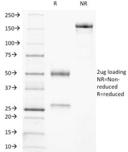
Purification Method
Protein A or G purified
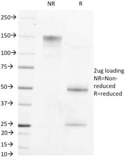
Test Specificity
Recognizes a phosphorylated protein of 228kDa, identified as nuclear mitotic apparatus protein (NuMA). Its epitope is resistant to phosphatases. NuMA is intra-nuclear protein and present in nucleus during interphase. At the onset of mitosis, it redistributes from the nucleus to two centrosomal structures that later will become part of the mitotic spindle pole. After anaphase, the protein redistributes from the spindle polar region into reforming nucleus. NuMA is an essential protein during mitosis for the terminal phases of chromosome separation and/or nuclear reassembly. Recently a study shows that NuMA is cleaved to a 180 to 200kDa during apoptosis. Chromosomal translocation of this gene with the RARA (retinoic acid receptor, alpha) gene on chromosome 17 has been detected in patients with acute promyelocytic leukemia.
Related Products
Description
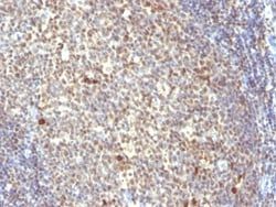
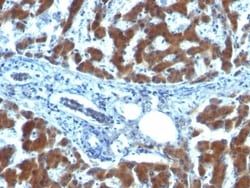
- Ensure accurate, reproducible results in Flow Cytometry, Immunohistochemistry (Paraffin), Immunofluorescence NuMA Monoclonal specifically detects NuMA in Human samples
- It is validated for Flow Cytometry, Immunohistochemistry, Immunocytochemistry/Immunofluorescence, Immunohistochemistry-Paraffin, Immunofluorescence.
Compare Similar Items
Show Difference
Antigen: NuMA
Concentration: 0.2 mg/mL
Applications: Flow Cytometry, Immunohistochemistry (Paraffin), Immunofluorescence
Conjugate: Unconjugated
Host Species: Mouse
Research Discipline: Breast Cancer, Cell Biology, Cell Cycle and Replication, Cellular Markers, Mitotic Regulators
Formulation: 10mM PBS and 0.05% BSA with 0.05% Sodium Azide
Gene ID (Entrez): 4926
Immunogen: Colon carcinoma 174T cells
Primary or Secondary: Primary
Content And Storage: Store at 4C.
Molecular Weight of Antigen: 228 kDa
Clone: SPM300
Dilution: Flow Cytometry 0.5 - 1 ug/million cells in 0.1 ml, Immunohistochemistry-Paraffin 0.5 - 1.0 ug/ml, Immunofluorescence 0.5 - 1.0 ug/ml
Classification: Monoclonal
Form: Purified
Regulatory Status: RUO
Target Species: Human
Gene Alias: centrophilin stabilizes mitotic spindle in mitotic cells, nuclear mitotic apparatus protein 1, NUMA, NuMA protein, SP-H antigen, structural nuclear protein
Gene Symbols: NUMA1
Isotype: IgM κ
Purification Method: Protein A or G purified
Test Specificity: Recognizes a phosphorylated protein of 228kDa, identified as nuclear mitotic apparatus protein (NuMA). Its epitope is resistant to phosphatases. NuMA is intra-nuclear protein and present in nucleus during interphase. At the onset of mitosis, it redistributes from the nucleus to two centrosomal structures that later will become part of the mitotic spindle pole. After anaphase, the protein redistributes from the spindle polar region into reforming nucleus. NuMA is an essential protein during mitosis for the terminal phases of chromosome separation and/or nuclear reassembly. Recently a study shows that NuMA is cleaved to a 180 to 200kDa during apoptosis. Chromosomal translocation of this gene with the RARA (retinoic acid receptor, alpha) gene on chromosome 17 has been detected in patients with acute promyelocytic leukemia.
Antigen: __
Concentration: __
Applications: __
Conjugate: __
Host Species: __
Research Discipline: __
Formulation: __
Gene ID (Entrez): __
Immunogen: __
Primary or Secondary: __
Content And Storage: __
Molecular Weight of Antigen: __
Clone: __
Dilution: __
Classification: __
Form: __
Regulatory Status: __
Target Species: __
Gene Alias: __
Gene Symbols: __
Isotype: __
Purification Method: __
Test Specificity: __
Antigen: Retinol Binding Protein RBP
Concentration: 0.2mg/mL
Applications: Immunohistochemistry (Paraffin), Immunofluorescence
Conjugate: Unconjugated
Host Species: Mouse
Research Discipline: __
Formulation: 10mM PBS and 0.05% BSA with 0.05% Sodium Azide
Gene ID (Entrez): 5947
Immunogen: Recombinant human retinol binding protein-1 (RBP1)
Primary or Secondary: Primary
Content And Storage: Store at 4C.
Molecular Weight of Antigen: __
Clone: RBP/872
Dilution: Immunohistochemistry-Paraffin 1 - 2 ug/ml, Immunofluorescence 1 - 2 ug/ml
Classification: Monoclonal
Form: Purified
Regulatory Status: RUO
Target Species: Human, Mouse, Rat, Goat, Monkey, Rabbit
Gene Alias: C, Cellular retinol-binding protein, CRABP-I, CRBP1CRBP-I, CRBPCellular retinol-binding protein I, CRBPI, RBPC, retinol binding protein 1, cellular, retinol-binding protein 1, retinol-binding protein 1, cellular
Gene Symbols: RBP1
Isotype: IgG1 κ
Purification Method: Protein A or G purified
Test Specificity: Recognizes a protein of 21kDa-25kDa, identified as retinol binding protein-1 (RBP1). This protein belongs to the lipocalin family and is the specific carrier for retinol (vitamin A alcohol) in the blood. It delivers retinol from the liver stores to the peripheral tissues. In plasma, the RBP-retinol complex interacts with transthyretin, which prevents its loss by filtration through the kidney glomeruli. A deficiency of vitamin A blocks secretion of the binding protein post-transnationally and results in defective delivery and supply to the epidermal cells.