alpha-Fetoprotein/AFP Antibody (MBS-12), Novus Biologicals™
Mouse Monoclonal Antibody
Manufacturer: Fischer Scientific
The price for this product is unavailable. Please request a quote
Antigen
alpha-Fetoprotein/AFP
Concentration
0.2mg/mL
Applications
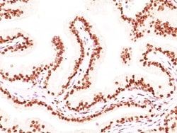
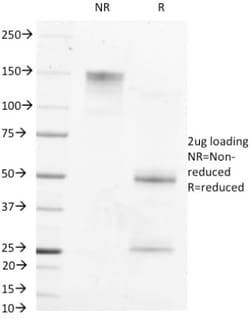
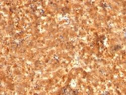
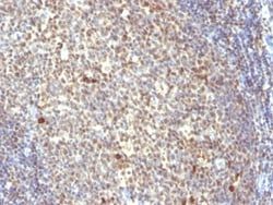
Flow Cytometry, Immunohistochemistry (Paraffin), SDS-Page, Immunofluorescence
Conjugate
Unconjugated
Host Species
Mouse
Research Discipline
Cancer, Cancer Stem Cells, GPCR, Stem Cell Markers, Tumor Biomarkers
Formulation
10mM PBS and 0.05% BSA with 0.05% Sodium Azide
Gene Alias
Alpha-1-fetoprotein, Alpha-fetoglobulin, alpha-fetoprotein, FETA, HP, HPAFP
Gene Symbols
AFP
Isotype
IgG1 κ
Purification Method
Protein A or G purified
Test Specificity
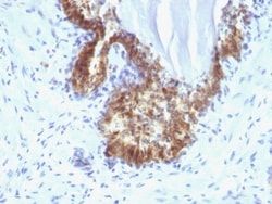
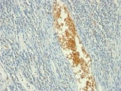
It recognizes an oncofetal glycoprotein with a single chain of 70kDa, which is identified as alpha fetoprotein (AFP). This MAb is highly specific to AFP and shows no cross-reaction with other oncofetal antigens or serum albumin. The yolk sac and the liver produce AFP during fetal life. AFP expression in adults is often associated with hepatoma or teratoma. However, hereditary persistence of alpha-fetoprotein may also be found in individuals with no obvious pathology. The protein is thought to be the fetal counterpart of serum albumin, and the AFP and albumin genes are present in tandem in the same transcriptional orientation on chromosome 4. AFP is found in monomeric as well as dimeric and trimeric forms, and binds copper, nickel, fatty acids and bilirubin. The level of AFP in amniotic fluid is used to measure renal loss of protein to screen for spinal bifida and anencephaly.
Clone
MBS-12
Dilution
Flow Cytometry 0.5 - 1 ug/million cells in 0.1 ml, Immunohistochemistry-Paraffin 1 - 2 ug/ml, SDS-Page, Immunofluorescence 0.5 - 1.0 ug/ml
Classification
Monoclonal
Form
Purified
Regulatory Status
RUO
Target Species
Human
Gene Accession No.
P02771
Gene ID (Entrez)
174
Immunogen
Recombinant full-length human Alpha fetoprotein
Primary or Secondary
Primary
Content And Storage
Store at 4C.
Molecular Weight of Antigen
70 kDa
Related Products

Description
- Ensure accurate, reproducible results in Flow Cytometry, Immunohistochemistry (Paraffin), Immunofluorescence alpha-Fetoprotein/AFP Monoclonal specifically detects alpha-Fetoprotein/AFP in Human samples
- It is validated for Flow Cytometry, Immunohistochemistry, Immunocytochemistry/Immunofluorescence, Immunohistochemistry-Paraffin, Immunofluorescence.
Compare Similar Items
Show Difference
Antigen: alpha-Fetoprotein/AFP
Concentration: 0.2mg/mL
Applications: Flow Cytometry, Immunohistochemistry (Paraffin), SDS-Page, Immunofluorescence
Conjugate: Unconjugated
Host Species: Mouse
Research Discipline: Cancer, Cancer Stem Cells, GPCR, Stem Cell Markers, Tumor Biomarkers
Formulation: 10mM PBS and 0.05% BSA with 0.05% Sodium Azide
Gene Alias: Alpha-1-fetoprotein, Alpha-fetoglobulin, alpha-fetoprotein, FETA, HP, HPAFP
Gene Symbols: AFP
Isotype: IgG1 κ
Purification Method: Protein A or G purified
Test Specificity: It recognizes an oncofetal glycoprotein with a single chain of 70kDa, which is identified as alpha fetoprotein (AFP). This MAb is highly specific to AFP and shows no cross-reaction with other oncofetal antigens or serum albumin. The yolk sac and the liver produce AFP during fetal life. AFP expression in adults is often associated with hepatoma or teratoma. However, hereditary persistence of alpha-fetoprotein may also be found in individuals with no obvious pathology. The protein is thought to be the fetal counterpart of serum albumin, and the AFP and albumin genes are present in tandem in the same transcriptional orientation on chromosome 4. AFP is found in monomeric as well as dimeric and trimeric forms, and binds copper, nickel, fatty acids and bilirubin. The level of AFP in amniotic fluid is used to measure renal loss of protein to screen for spinal bifida and anencephaly.
Clone: MBS-12
Dilution: Flow Cytometry 0.5 - 1 ug/million cells in 0.1 ml, Immunohistochemistry-Paraffin 1 - 2 ug/ml, SDS-Page, Immunofluorescence 0.5 - 1.0 ug/ml
Classification: Monoclonal
Form: Purified
Regulatory Status: RUO
Target Species: Human
Gene Accession No.: P02771
Gene ID (Entrez): 174
Immunogen: Recombinant full-length human Alpha fetoprotein
Primary or Secondary: Primary
Content And Storage: Store at 4C.
Molecular Weight of Antigen: 70 kDa

Antigen: Aminopeptidase N/CD13
Concentration: 0.2 mg/mL
Applications: Flow Cytometry, Immunohistochemistry (Paraffin), Immunofluorescence
Conjugate: Unconjugated
Host Species: Mouse
Research Discipline: Angiogenesis, Cellular Markers, Hematopoietic Stem Cell Markers, Immunology, Mesenchymal Stem Cell Markers, Stem Cell Markers
Formulation: 10mM PBS and 0.05% BSA with 0.05% Sodium Azide
Gene Alias: alanyl (membrane) aminopeptidase, Alanyl aminopeptidase, Aminopeptidase M, aminopeptidase N, AP-M, AP-N, CD13 antigen, CD13APN, EC 3.4.11, EC 3.4.11.2, gp150, LAP1, Microsomal aminopeptidase, Myeloid plasma membrane glycoprotein CD13, P150, PEPNhAPN
Gene Symbols: ANPEP
Isotype: IgG1 κ
Purification Method: Protein A or G purified
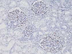
Test Specificity: This MAb recognizes an extracellular epitope of an integral membrane glycoprotein of 150kDa, identified as CD13. This antigen is present on most cells of myeloid origin including granulocytes, monocytes, mast cells, and GM-progenitor cells. It is also expressed by the majority of AML, CML in myeloid blast crisis, and in a smaller fraction of lymphoid leukemias. It is absent from normal lymphocytes, platelets and erythrocytes. CD13 is also present on fibroblasts; endothelial cells, epithelial cells from renal proximal tubules and intestinal brush border, bone marrow stromal cells, osteoclasts, and cells lining bile duct canaliculi. CD13 is identical to aminopeptidase N (APN), a prominent membrane-bound metalloprotease present on the surface of intestinal brush border and renal tubules. CD13 plays a role in metabolism of biologically active peptides, in phagocytosis, and in bactericidal/tumoricidal activities. It also serves as a receptor for human coronaviruses (HCV). The lineage-restricted pattern of expression of CD13 within the hemopoietic compartment suggests that it may be important in myeloid cell differentiation.
Clone: APN/514
Dilution: Flow Cytometry 0.5 - 1 ug/million cells in 0.1 ml, Immunohistochemistry-Paraffin 0.5 - 1.0 ug/ml, Immunofluorescence 1 - 2 ug/ml
Classification: Monoclonal
Form: Purified
Regulatory Status: RUO
Target Species: Human
Gene Accession No.: P15144, P15144
Gene ID (Entrez): 290
Immunogen: Recombinant human CD13 protein
Primary or Secondary: Primary
Content And Storage: Store at 4C.
Molecular Weight of Antigen: 150 kDa
Antigen: AMPD3
Concentration: 0.2mg/mL
Applications: Flow Cytometry, Immunohistochemistry (Paraffin), Immunofluorescence, Immunocytochemistry
Conjugate: Unconjugated
Host Species: Mouse
Research Discipline: __
Formulation: 10mM PBS and 0.05% BSA with 0.05% Sodium Azide
Gene Alias: adenosine monophosphate deaminase (isoform E), adenosine monophosphate deaminase 3, AMP aminohydrolase, AMP deaminase 3, AMP deaminase isoform E, EC 3.5.4.6, Erythrocyte AMP deaminase, erythrocyte type AMP deaminase, erythrocyte-specific AMP deaminase, myoadenylate deaminase
Gene Symbols: AMPD3
Isotype: IgG2b κ
Purification Method: Protein A or G purified
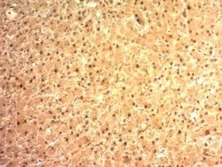
Test Specificity: It recognizes a protein of ∼90kDa, which is identified as Adenosine Monophosphate Deaminase, isoform E (AMPD3). It has 767 amino acids and is assigned an EC 3.5.4.6. It is a highly regulated enzyme that catalyzes the hydrolytic deamination of adenosine monophosphate to inosine monophosphate, a branch point in the adenylate catabolic pathway. AMPD3 gene encodes the erythrocyte (E) isoforms, whereas other family members encode isoforms that predominate in muscle (M) and liver (L) cells. This MAb shows reactivity with cells of the erythroid lineage at all stages of maturation in the peripheral blood, bone marrow, and fetal liver. Non-erythroid lineages are negative by flow cytometry. This MAb is useful in the diagnosis of erythroleukemia, identification of bone marrow erythroid precursors, gating erythroid nucleated precursor cells from malignant cells in bone marrow specimens.
Clone: AMPD3/901
Dilution: Flow Cytometry 1 - 2 ug/million cells in 0.1ml, Immunohistochemistry-Paraffin 2 - 4 ug/ml, Immunofluorescence 0.5 - 1.0 ug/ml, Immunocytochemistry 1 - 2.0 ug/ml
Classification: Monoclonal
Form: Purified
Regulatory Status: RUO
Target Species: Human
Gene Accession No.: Q01432
Gene ID (Entrez): 272
Immunogen: Recombinant full-length human AMDP3 protein
Primary or Secondary: Primary
Content And Storage: Store at 4C.
Molecular Weight of Antigen: __