AMPD3 Antibody (AMPD3/901), Novus Biologicals™
Mouse Monoclonal Antibody
Manufacturer: Fischer Scientific
The price for this product is unavailable. Please request a quote
Antigen
AMPD3
Concentration
0.2mg/mL
Applications
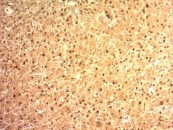
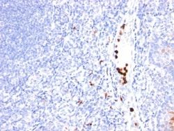
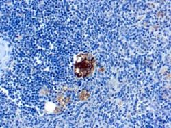
Flow Cytometry, Immunohistochemistry (Paraffin), Immunofluorescence, Immunocytochemistry
Conjugate
Unconjugated
Host Species
Mouse
Target Species
Human
Gene Accession No.
Q01432
Gene ID (Entrez)
272
Immunogen
Recombinant full-length human AMDP3 protein
Primary or Secondary
Primary
Content And Storage
Store at 4C.
Clone
AMPD3/901
Dilution
Flow Cytometry 1 - 2 ug/million cells in 0.1ml, Immunohistochemistry-Paraffin 2 - 4 ug/ml, Immunofluorescence 0.5 - 1.0 ug/ml, Immunocytochemistry 1 - 2.0 ug/ml
Classification
Monoclonal
Form
Purified
Regulatory Status
RUO
Formulation
10mM PBS and 0.05% BSA with 0.05% Sodium Azide
Gene Alias
adenosine monophosphate deaminase (isoform E), adenosine monophosphate deaminase 3, AMP aminohydrolase, AMP deaminase 3, AMP deaminase isoform E, EC 3.5.4.6, Erythrocyte AMP deaminase, erythrocyte type AMP deaminase, erythrocyte-specific AMP deaminase, myoadenylate deaminase
Gene Symbols
AMPD3
Isotype
IgG2b κ
Purification Method
Protein A or G purified
Test Specificity
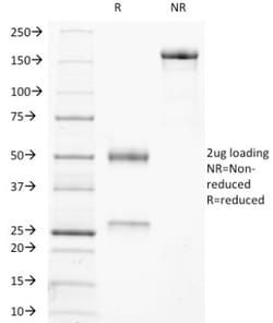
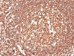
It recognizes a protein of ∼90kDa, which is identified as Adenosine Monophosphate Deaminase, isoform E (AMPD3). It has 767 amino acids and is assigned an EC 3.5.4.6. It is a highly regulated enzyme that catalyzes the hydrolytic deamination of adenosine monophosphate to inosine monophosphate, a branch point in the adenylate catabolic pathway. AMPD3 gene encodes the erythrocyte (E) isoforms, whereas other family members encode isoforms that predominate in muscle (M) and liver (L) cells. This MAb shows reactivity with cells of the erythroid lineage at all stages of maturation in the peripheral blood, bone marrow, and fetal liver. Non-erythroid lineages are negative by flow cytometry. This MAb is useful in the diagnosis of erythroleukemia, identification of bone marrow erythroid precursors, gating erythroid nucleated precursor cells from malignant cells in bone marrow specimens.
Related Products


Description
- Ensure accurate, reproducible results in Flow Cytometry, Immunohistochemistry (Paraffin), Immunocytochemistry, Immunofluorescence AMPD3 Monoclonal specifically detects AMPD3 in Human samples
- It is validated for Immunohistochemistry, Immunohistochemistry-Paraffin.
Compare Similar Items
Show Difference
Antigen: AMPD3
Concentration: 0.2mg/mL
Applications: Flow Cytometry, Immunohistochemistry (Paraffin), Immunofluorescence, Immunocytochemistry
Conjugate: Unconjugated
Host Species: Mouse
Target Species: Human
Gene Accession No.: Q01432
Gene ID (Entrez): 272
Immunogen: Recombinant full-length human AMDP3 protein
Primary or Secondary: Primary
Content And Storage: Store at 4C.
Clone: AMPD3/901
Dilution: Flow Cytometry 1 - 2 ug/million cells in 0.1ml, Immunohistochemistry-Paraffin 2 - 4 ug/ml, Immunofluorescence 0.5 - 1.0 ug/ml, Immunocytochemistry 1 - 2.0 ug/ml
Classification: Monoclonal
Form: Purified
Regulatory Status: RUO
Formulation: 10mM PBS and 0.05% BSA with 0.05% Sodium Azide
Gene Alias: adenosine monophosphate deaminase (isoform E), adenosine monophosphate deaminase 3, AMP aminohydrolase, AMP deaminase 3, AMP deaminase isoform E, EC 3.5.4.6, Erythrocyte AMP deaminase, erythrocyte type AMP deaminase, erythrocyte-specific AMP deaminase, myoadenylate deaminase
Gene Symbols: AMPD3
Isotype: IgG2b κ
Purification Method: Protein A or G purified
Test Specificity: It recognizes a protein of ∼90kDa, which is identified as Adenosine Monophosphate Deaminase, isoform E (AMPD3). It has 767 amino acids and is assigned an EC 3.5.4.6. It is a highly regulated enzyme that catalyzes the hydrolytic deamination of adenosine monophosphate to inosine monophosphate, a branch point in the adenylate catabolic pathway. AMPD3 gene encodes the erythrocyte (E) isoforms, whereas other family members encode isoforms that predominate in muscle (M) and liver (L) cells. This MAb shows reactivity with cells of the erythroid lineage at all stages of maturation in the peripheral blood, bone marrow, and fetal liver. Non-erythroid lineages are negative by flow cytometry. This MAb is useful in the diagnosis of erythroleukemia, identification of bone marrow erythroid precursors, gating erythroid nucleated precursor cells from malignant cells in bone marrow specimens.
Antigen: Androgen R/NR3C4
Concentration: 0.2mg/mL
Applications: Immunohistochemistry (Paraffin), Immunofluorescence
Conjugate: Unconjugated
Host Species: Mouse
Target Species: Human, Mouse (Negative)
Gene Accession No.: P10275
Gene ID (Entrez): 367
Immunogen: A synthetic peptide, aa 299-315, (STEDTAEYSPFKGGYTK) of human AR (AR441); Recombinant human DHTR protein (DHTR/882)
Primary or Secondary: Primary
Content And Storage: Store at 4C.
Clone: AR441 + DHTR/882
Dilution: Immunohistochemistry-Paraffin 0.5 - 1.0 ug/ml, Immunofluorescence 0.5 - 1.0 ug/ml
Classification: Monoclonal
Form: Purified
Regulatory Status: RUO
Formulation: 1.0mM PBS and 0.05% BSA with 0.05% Sodium Azide
Gene Alias: AIS, androgen receptor, DHTRTFM, Dihydrotestosterone receptorHYSP1, HUMARA, NR3C4KD, Nuclear receptor subfamily 3 group C member 4, SMAX1SBMA, spinal and bulbar muscular atrophy
Gene Symbols: AR
Isotype: IgG
Purification Method: Protein A or G purified
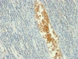
Test Specificity: Recognizes a protein of 110kDa, which is identified as androgen receptor (AR). It reacts with full length, and the newly described A form of the receptor. It does not cross react with estrogen, progesterone, or glucocorticoid receptors. The expression of AR is reportedly inversely correlated with histologic grade i.e. well differentiated prostate tumors show higher expression than the poorly differentiated tumors. In prostate cancer, AR has been proposed, as a marker of hormone-responsiveness and thus it may be useful in identifying patients likely to benefit from anti-androgen therapy. Anti-androgen receptor has been useful clinically in differentiating morpheaform basal cell carcinoma (mBCC) from desmoplastic trichoepithelioma (DTE) in the skin.This MAb is superb for staining of formalin/paraffin tissues.
Antigen: Androgen R/NR3C4
Concentration: 0.2mg/mL
Applications: Flow Cytometry, Immunohistochemistry (Paraffin), Immunofluorescence
Conjugate: Unconjugated
Host Species: Mouse
Target Species: Human, Mouse (Negative)
Gene Accession No.: P10275
Gene ID (Entrez): 367
Immunogen: Recombinant full-length human DHTR protein
Primary or Secondary: Primary
Content And Storage: Store at 4C.
Clone: DHTR/882
Dilution: Flow Cytometry 0.5 - 1 ug/million cells in 0.1 ml, Immunohistochemistry-Paraffin 0.5 - 1.0 ug/ml, Immunofluorescence 0.5 - 1.0 ug/ml
Classification: Monoclonal
Form: Purified
Regulatory Status: RUO
Formulation: 1.0mM PBS and 0.05% BSA with 0.05% Sodium Azide
Gene Alias: AIS, androgen receptor, DHTRTFM, Dihydrotestosterone receptorHYSP1, HUMARA, NR3C4KD, Nuclear receptor subfamily 3 group C member 4, SMAX1SBMA, spinal and bulbar muscular atrophy
Gene Symbols: AR
Isotype: IgG1 κ
Purification Method: Protein A or G purified
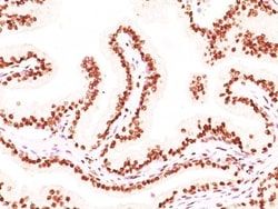
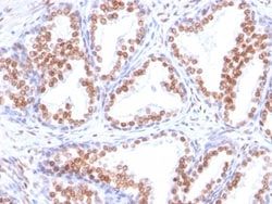
Test Specificity: Recognizes a protein of 110kDa, which is identified as androgen receptor (AR). It reacts with full length, and the newly described A form of the receptor. It does not cross react with estrogen, progesterone, or glucocorticoid receptors. The expression of AR is reportedly inversely correlated with histologic grade i.e. well differentiated prostate tumors show higher expression than the poorly differentiated tumors. In prostate cancer, AR has been proposed, as a marker of hormone-responsiveness and thus it may be useful in identifying patients likely to benefit from anti-androgen therapy. Anti-androgen receptor has been useful clinically in differentiating morpheaform basal cell carcinoma (mBCC) from desmoplastic trichoepithelioma (DTE) in the skin.This MAb is superb for staining of formalin/paraffin tissues.