MITF Antibody (C5/D5), Novus Biologicals™
Mouse Monoclonal Antibody
Manufacturer: Fischer Scientific
The price for this product is unavailable. Please request a quote
Antigen
MITF
Concentration
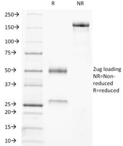
0.2mg/mL
Applications
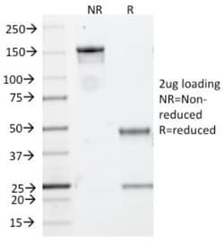
Flow Cytometry, Immunohistochemistry (Paraffin), SDS-Page, Immunofluorescence
Conjugate
Unconjugated
Host Species
Mouse
Target Species
Human, Mouse (Negative), Rat (Negative)
Gene Alias
BHLHE32, bHLHe32MI, Class E basic helix-loop-helix protein 32, microphthalmia-associated transcription factor, Waardenburg syndrome, type 2A, WS2A
Gene Symbols
MITF
Isotype
IgG1 κ
Purification Method
Protein A or G purified
Test Specificity
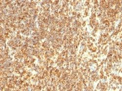
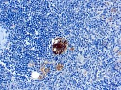
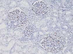
MITF (microphthalmia transcription factor) is a basic helix-loop-helix-leucine-zipper (bHLH-Zip) transcription factor that regulates the development and survival of melanocytes and retinal pigment epithelium, and also is involved in transcription of pigmentation enzyme genes such as tyrosinase TRP1 and TRP2. MITF has been shown to be phosphorylated by MAP kinase in response to c-kit activation, resulting in upregulation of MITF transcriptional activity. Mutations of the MITF gene are associated with the autosomal dominant hereditary deafness and pigmentation condition, Waardenburg Syndrome type 2A. Multiple isoforms of MITF exist, including MITF-A, MITF-B, MITF-C, MITF-H, and MITF-M, which differ in the amino-terminal domain and in their expression patterns. The MITF-M isoform is restricted to the melanocyte cell lineage. Anti-MITF, D5, recognizes a nuclear protein, which is expressed in the majority of primary and metastatic epithelioid malignant melanomas as well as in normal melanoc
Clone
D5
Dilution
Flow Cytometry 0.5 - 1 ug/million cells in 0.1 ml, Immunohistochemistry-Paraffin 0.5 - 1.0 ug/ml, SDS-Page, Immunofluorescence 0.5 - 1.0 ug/ml
Classification
Monoclonal
Form
Purified
Regulatory Status
RUO
Formulation
10mM PBS and 0.05% BSA with 0.05% Sodium Azide
Gene ID (Entrez)
4286
Immunogen
NH2 terminus fragment of human Mi protein
Primary or Secondary
Primary
Content And Storage
Store at 4C.
Related Products

Description
- Ensure accurate, reproducible results in Flow Cytometry, Immunohistochemistry (Paraffin), Immunofluorescence MITF Monoclonal specifically detects MITF in Human, Mouse (Negative), Rat (Negative) samples
- It is validated for Immunohistochemistry, Immunohistochemistry-Paraffin.
Compare Similar Items
Show Difference
Antigen: MITF
Concentration: 0.2mg/mL
Applications: Flow Cytometry, Immunohistochemistry (Paraffin), SDS-Page, Immunofluorescence
Conjugate: Unconjugated
Host Species: Mouse
Target Species: Human, Mouse (Negative), Rat (Negative)
Gene Alias: BHLHE32, bHLHe32MI, Class E basic helix-loop-helix protein 32, microphthalmia-associated transcription factor, Waardenburg syndrome, type 2A, WS2A
Gene Symbols: MITF
Isotype: IgG1 κ
Purification Method: Protein A or G purified
Test Specificity: MITF (microphthalmia transcription factor) is a basic helix-loop-helix-leucine-zipper (bHLH-Zip) transcription factor that regulates the development and survival of melanocytes and retinal pigment epithelium, and also is involved in transcription of pigmentation enzyme genes such as tyrosinase TRP1 and TRP2. MITF has been shown to be phosphorylated by MAP kinase in response to c-kit activation, resulting in upregulation of MITF transcriptional activity. Mutations of the MITF gene are associated with the autosomal dominant hereditary deafness and pigmentation condition, Waardenburg Syndrome type 2A. Multiple isoforms of MITF exist, including MITF-A, MITF-B, MITF-C, MITF-H, and MITF-M, which differ in the amino-terminal domain and in their expression patterns. The MITF-M isoform is restricted to the melanocyte cell lineage. Anti-MITF, D5, recognizes a nuclear protein, which is expressed in the majority of primary and metastatic epithelioid malignant melanomas as well as in normal melanoc
Clone: D5
Dilution: Flow Cytometry 0.5 - 1 ug/million cells in 0.1 ml, Immunohistochemistry-Paraffin 0.5 - 1.0 ug/ml, SDS-Page, Immunofluorescence 0.5 - 1.0 ug/ml
Classification: Monoclonal
Form: Purified
Regulatory Status: RUO
Formulation: 10mM PBS and 0.05% BSA with 0.05% Sodium Azide
Gene ID (Entrez): 4286
Immunogen: NH2 terminus fragment of human Mi protein
Primary or Secondary: Primary
Content And Storage: Store at 4C.
Antigen: Moesin
Concentration: 0.2mg/mL
Applications: Western Blot, Flow Cytometry, Immunohistochemistry (Paraffin), Immunofluorescence
Conjugate: Unconjugated
Host Species: Mouse
Target Species: Human, Rat (Negative)
Gene Alias: Membrane-organizing extension spike protein, moesin
Gene Symbols: MSN
Isotype: IgG1 κ
Purification Method: Protein A or G purified
Test Specificity: Recognizes 78kDa moesin protein. Moesin, a member of the talin-4.1 superfamily, is a linking protein of the sub-membranous actin cytoskeleton. It is expressed in variable amounts in cells of different phenotypes such as macrophages, lymphocytes, fibroblastic, endothelial, epithelial, and neuronal cell lines but not in blood cells. The ERM proteins, ezrin, radixin, and moesin are involved in a variety of cellular functions, such as cell adhesion, migration, and the organization of cell surface structures, and are highly homologous, both in protein sequence and in functional activity, with merlin/schwannomin, a neurofibromatosis-2-associated tumor-suppressor protein. Cell lines of epithelial and mesothelial origin contain both moesin and radixin whereas cells of endothelial and lymphoid origin express moesin.
Clone: MSN/492
Dilution: Western Blot 0.5 - 1.0 ug/ml, Flow Cytometry 0.5 - 1 ug/million cells in 0.1 ml, Immunohistochemistry-Paraffin 0.5 - 1.0 ug/ml, Immunofluorescence 0.5 - 1.0 ug/ml
Classification: Monoclonal
Form: Purified
Regulatory Status: RUO
Formulation: 10mM PBS and 0.05% BSA with 0.05% Sodium Azide
Gene ID (Entrez): 4478
Immunogen: Recombinant full-length human Moesin protein
Primary or Secondary: Primary
Content And Storage: Store at 4C.
Antigen: CD20
Concentration: 0.2mg/mL
Applications: Flow Cytometry, Immunohistochemistry (Paraffin), SDS-Page, Immunofluorescence
Conjugate: Unconjugated
Host Species: Mouse
Target Species: Human
Gene Alias: B1, B-lymphocyte antigen CD20, B-lymphocyte cell-surface antigen B1, B-lymphocyte surface antigen B1, Bp35MGC3969, CD20 antigen, CD20 receptor, CD20S7, CVID5, LEU-16, Leukocyte surface antigen Leu-16, Membrane-spanning 4-domains subfamily A member 1, membrane-spanning 4-domains, subfamily A, member 1, MS4A2
Gene Symbols: MS4A1
Isotype: IgG2a κ
Purification Method: Protein A or G purified
Test Specificity: Recognizes a protein of 30-33kDa, which is identified as CD20. It is a non-Ig differentiation antigen of B-cells and its expression is restricted to normal and neoplastic B-cells, being absent from all other leukocytes and tissues. CD20 is expressed by pre B-cells and persists during all stages of B-cell maturation but is lost upon terminal differentiation into plasma cells. This MAb can be used for immunophenotyping of leukemia and malignant cells, B lymphocyte detection in peripheral blood and B cell localization in tissues. It reacts with the majority of B-cells present in peripheral blood and lymphoid tissues and their derived lymphomas. In lymphoid tissue, germinal center blasts and B-immunoblasts are particularly reactive. It is a reliable antibody for ascribing a B-cell phenotype in known lymphoid tissues. Rarely, CD20-positive T-cell lymphomas have been reported. Reactivity has also been noted with Reed-Sternberg cells in cases of Hodgkin s disease, particularly of lymphocyte p
Clone: IGEL/773
Dilution: Flow Cytometry 0.5 - 1 ug/million cells in 0.1 ml, Immunohistochemistry-Paraffin 0.5 - 1.0 ug/ml, SDS-Page, Immunofluorescence 0.5 - 1.0 ug/ml
Classification: Monoclonal
Form: Purified
Regulatory Status: RUO
Formulation: 10mM PBS and 0.05% BSA with 0.05% Sodium Azide
Gene ID (Entrez): 931
Immunogen: Recombinant human MS4A1 protein
Primary or Secondary: Primary
Content And Storage: Store at 4C.