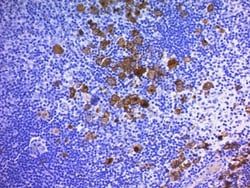
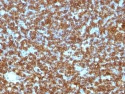
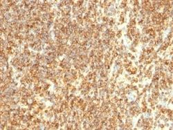
CD20 Antibody (SPM494), Novus Biologicals™
Mouse Monoclonal Antibody
Manufacturer: Fischer Scientific
The price for this product is unavailable. Please request a quote
Antigen
CD20
Concentration
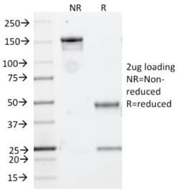
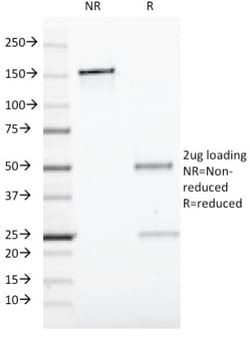
0.2mg/mL
Applications
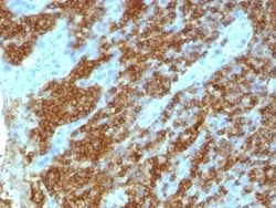
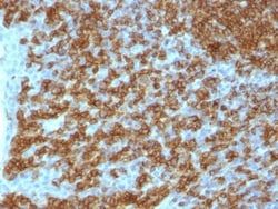
Flow Cytometry, Immunohistochemistry (Paraffin), Immunofluorescence
Conjugate
Unconjugated
Host Species
Mouse
Research Discipline
Adaptive Immunity, Cancer Stem Cells, Cell Biology, Cytokine Research, Immunology, Signal Transduction, Stem Cell Markers, Tumor Biomarkers
Formulation
No buffer with 0.05% Sodium Azide
Gene Alias
B1, B-lymphocyte antigen CD20, B-lymphocyte cell-surface antigen B1, B-lymphocyte surface antigen B1, Bp35MGC3969, CD20 antigen, CD20 receptor, CD20S7, CVID5, LEU-16, Leukocyte surface antigen Leu-16, Membrane-spanning 4-domains subfamily A member 1, membrane-spanning 4-domains, subfamily A, member 1, MS4A2
Gene Symbols
MS4A1
Isotype
IgG2a κ
Purification Method
Tissue culture supernatant
Test Specificity
Recognizes a protein of 30-33kDa, which is identified as CD20. Its epitope is located in the cytoplasmic domain of CD20 and was, therefore, ascribed as CD20cy in the 5th Workshop. CD20 is a non-Ig differentiation antigen of B-cells and its expression is restricted to normal and neoplastic B-cells, being absent from all other leukocytes and tissues. CD20 is expressed by pre B-cells and persists during all stages of B-cell maturation but is lost upon terminal differentiation into plasma cells. This MAb can be used for immunophenotyping of leukemia and malignant cells, B lymphocyte detection in peripheral blood and B cell localization in tissues. It reacts with the majority of B-cells present in peripheral blood and lymphoid tissues and their derived lymphomas. In lymphoid tissue, germinal center blasts and B-immunoblasts are particularly reactive. It is a reliable antibody for ascribing a B-cell phenotype in known lymphoid tissues. Rarely, CD20-positive T-cell lymphomas have been reporte
Clone
SPM494
Dilution
Flow Cytometry 2 - 5ul/million cells in 0.1ml, Immunohistochemistry-Paraffin 1:100-1:200, Immunofluorescence 1:100-1:200
Classification
Monoclonal
Form
Supernatant
Regulatory Status
RUO
Target Species
Human, Baboon, Monkey, Bovine (Negative), Canine (Negative), Porcine (Negative), Rat (Negative)
Gene Accession No.
P11836
Gene ID (Entrez)
931
Immunogen
Human tonsil B cells
Primary or Secondary
Primary
Content And Storage
Store at 4C.
Molecular Weight of Antigen
35 kDa
Related Products
Description
- Ensure accurate, reproducible results in Flow Cytometry, Immunohistochemistry (Paraffin), Immunofluorescence CD20 Monoclonal specifically detects CD20 in Human, Baboon, Monkey, Bovine (Negative), Canine (Negative), Porcine (Negative), Rat (Negative) samples
- It is validated for Flow Cytometry, Immunohistochemistry, Immunocytochemistry/Immunofluorescence, Immunohistochemistry-Paraffin.
Compare Similar Items
Show Difference
Antigen: CD20
Concentration: 0.2mg/mL
Applications: Flow Cytometry, Immunohistochemistry (Paraffin), Immunofluorescence
Conjugate: Unconjugated
Host Species: Mouse
Research Discipline: Adaptive Immunity, Cancer Stem Cells, Cell Biology, Cytokine Research, Immunology, Signal Transduction, Stem Cell Markers, Tumor Biomarkers
Formulation: No buffer with 0.05% Sodium Azide
Gene Alias: B1, B-lymphocyte antigen CD20, B-lymphocyte cell-surface antigen B1, B-lymphocyte surface antigen B1, Bp35MGC3969, CD20 antigen, CD20 receptor, CD20S7, CVID5, LEU-16, Leukocyte surface antigen Leu-16, Membrane-spanning 4-domains subfamily A member 1, membrane-spanning 4-domains, subfamily A, member 1, MS4A2
Gene Symbols: MS4A1
Isotype: IgG2a κ
Purification Method: Tissue culture supernatant
Test Specificity: Recognizes a protein of 30-33kDa, which is identified as CD20. Its epitope is located in the cytoplasmic domain of CD20 and was, therefore, ascribed as CD20cy in the 5th Workshop. CD20 is a non-Ig differentiation antigen of B-cells and its expression is restricted to normal and neoplastic B-cells, being absent from all other leukocytes and tissues. CD20 is expressed by pre B-cells and persists during all stages of B-cell maturation but is lost upon terminal differentiation into plasma cells. This MAb can be used for immunophenotyping of leukemia and malignant cells, B lymphocyte detection in peripheral blood and B cell localization in tissues. It reacts with the majority of B-cells present in peripheral blood and lymphoid tissues and their derived lymphomas. In lymphoid tissue, germinal center blasts and B-immunoblasts are particularly reactive. It is a reliable antibody for ascribing a B-cell phenotype in known lymphoid tissues. Rarely, CD20-positive T-cell lymphomas have been reporte
Clone: SPM494
Dilution: Flow Cytometry 2 - 5ul/million cells in 0.1ml, Immunohistochemistry-Paraffin 1:100-1:200, Immunofluorescence 1:100-1:200
Classification: Monoclonal
Form: Supernatant
Regulatory Status: RUO
Target Species: Human, Baboon, Monkey, Bovine (Negative), Canine (Negative), Porcine (Negative), Rat (Negative)
Gene Accession No.: P11836
Gene ID (Entrez): 931
Immunogen: Human tonsil B cells
Primary or Secondary: Primary
Content And Storage: Store at 4C.
Molecular Weight of Antigen: 35 kDa
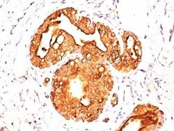
Antigen: MUC-1
Concentration: 0.2mg/mL
Applications: Flow Cytometry, Immunohistochemistry (Paraffin), Immunofluorescence
Conjugate: Unconjugated
Host Species: Mouse
Research Discipline: Cancer, Cellular Markers, Extracellular Matrix
Formulation: 10mM PBS and 0.05% BSA with 0.05% Sodium Azide
Gene Alias: Breast carcinoma-associated antigen DF3, Carcinoma-associated mucin, CD227, CD227 antigen, DF3 antigen, EMA, episialin, H23 antigen, H23AG, KL-6, MAM6, MUC-1, MUC1/ZD, mucin 1, cell surface associated, mucin 1, transmembrane, mucin-1, Peanut-reactive urinary mucin, PEMMUC-1/SEC, PEMT, Polymorphic epithelial mucin, PUMMUC-1/X, tumor associated epithelial mucin, Tumor-associated epithelial membrane antigen, Tumor-associated mucin
Gene Symbols: MUC1
Isotype: IgG1 κ
Purification Method: Protein A or G purified
Test Specificity: __
Clone: MUC1/955
Dilution: Flow Cytometry 0.5 - 1 ug/million cells in 0.1 ml, Immunohistochemistry-Paraffin 0.25 - 0.5 ug/ml, Immunofluorescence 1 - 2 ug/ml
Classification: Monoclonal
Form: Purified
Regulatory Status: RUO
Target Species: Human, Mouse
Gene Accession No.: P15941
Gene ID (Entrez): 4582
Immunogen: Human milk-fat globule membranes (HMFGM)
Primary or Secondary: Primary
Content And Storage: Store at 4C.
Molecular Weight of Antigen: __
