Tenascin C Antibody (T2H5), Novus Biologicals™
Mouse Monoclonal Antibody
Manufacturer: Fischer Scientific
The price for this product is unavailable. Please request a quote
Antigen
Tenascin C
Concentration
0.2mg/mL
Applications
Flow Cytometry, Immunohistochemistry (Paraffin), Immunofluorescence
Conjugate
Unconjugated
Host Species
Mouse
Research Discipline
Breast Cancer, Cancer, Extracellular Matrix, Neuroscience, Prostate Cancer
Formulation
1.0mM PBS and 0.05% BSA with 0.05% Sodium Azide
Gene Alias
150-225, Cytotactin, Glioma-associated-extracellular matrix antigen, GMEM, GP 150-225, Hexabrachion, hexabrachion (tenascin C, cytotactin), hexabrachion (tenascin), HXBcytotactin, JI, MGC167029, Myotendinous antigen, neuronectin, tenascin, tenascin C, Tenascin-C, tenascin-C isoform 14/AD1/16, TN-C, TNGP
Gene Symbols
TNC
Isotype
IgG1 κ
Purification Method
Protein A or G purified
Test Specificity
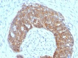
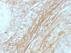
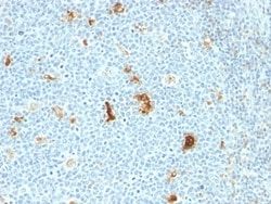
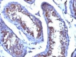
In Western blotting, it reacts with two bands of ∼MW of 210kDa and 300kDa, identified as two isoforms of Tenascin C. Specificity of this MAb is validated by sequential immunoprecipitation with a PAb against Tenascin C. Tenascin C is a multifunctional, disulfide-linked hexameric extracellular matrix glycoprotein expressed in association with mesenchymal epithelial interactions during development and in the neo-vasculature and stroma of undifferentiated tumors. In adults, it is restricted to certain epithelial-stromal interfaces and increases markedly in hyper-proliferative diseases and in stroma of many neoplasms, including gliomas, breast, squamous and lung carcinomas.
Clone
T2H5
Dilution
Flow Cytometry 0.5 - 1 ug/million cells in 0.1 ml, Immunohistochemistry-Paraffin 2 - 4 ug/ml, Immunofluorescence 0.5 - 1.0 ug/ml
Classification
Monoclonal
Form
Purified
Regulatory Status
RUO
Target Species
Human, Rat (Negative)
Gene Accession No.
P24821
Gene ID (Entrez)
3371
Immunogen
Human breast carcinoma
Primary or Secondary
Primary
Content And Storage
Store at 4C.
Related Products


Description
- Ensure accurate, reproducible results in Flow Cytometry, Immunohistochemistry (Paraffin), Immunofluorescence Tenascin C Monoclonal specifically detects Tenascin C in Human, Rat (Negative) samples
- It is validated for Immunohistochemistry, Immunohistochemistry-Paraffin.
Compare Similar Items
Show Difference
Antigen: Tenascin C
Concentration: 0.2mg/mL
Applications: Flow Cytometry, Immunohistochemistry (Paraffin), Immunofluorescence
Conjugate: Unconjugated
Host Species: Mouse
Research Discipline: Breast Cancer, Cancer, Extracellular Matrix, Neuroscience, Prostate Cancer
Formulation: 1.0mM PBS and 0.05% BSA with 0.05% Sodium Azide
Gene Alias: 150-225, Cytotactin, Glioma-associated-extracellular matrix antigen, GMEM, GP 150-225, Hexabrachion, hexabrachion (tenascin C, cytotactin), hexabrachion (tenascin), HXBcytotactin, JI, MGC167029, Myotendinous antigen, neuronectin, tenascin, tenascin C, Tenascin-C, tenascin-C isoform 14/AD1/16, TN-C, TNGP
Gene Symbols: TNC
Isotype: IgG1 κ
Purification Method: Protein A or G purified
Test Specificity: In Western blotting, it reacts with two bands of ∼MW of 210kDa and 300kDa, identified as two isoforms of Tenascin C. Specificity of this MAb is validated by sequential immunoprecipitation with a PAb against Tenascin C. Tenascin C is a multifunctional, disulfide-linked hexameric extracellular matrix glycoprotein expressed in association with mesenchymal epithelial interactions during development and in the neo-vasculature and stroma of undifferentiated tumors. In adults, it is restricted to certain epithelial-stromal interfaces and increases markedly in hyper-proliferative diseases and in stroma of many neoplasms, including gliomas, breast, squamous and lung carcinomas.
Clone: T2H5
Dilution: Flow Cytometry 0.5 - 1 ug/million cells in 0.1 ml, Immunohistochemistry-Paraffin 2 - 4 ug/ml, Immunofluorescence 0.5 - 1.0 ug/ml
Classification: Monoclonal
Form: Purified
Regulatory Status: RUO
Target Species: Human, Rat (Negative)
Gene Accession No.: P24821
Gene ID (Entrez): 3371
Immunogen: Human breast carcinoma
Primary or Secondary: Primary
Content And Storage: Store at 4C.
Antigen: Testosterone
Concentration: 0.2 mg/mL
Applications: Immunohistochemistry (Paraffin), Radioimmune Assays (RIA), SDS-Page
Conjugate: Unconjugated
Host Species: Mouse
Research Discipline: __
Formulation: 10mM PBS and 0.05% BSA with 0.05% Sodium Azide
Gene Alias: __
Gene Symbols: __
Isotype: IgG1 κ
Purification Method: Protein A or G purified
Test Specificity: This MAb is highly specific to testosterone. Its affinity constant for testosterone is ∼1010M-1. In competitive binding immunoassay, it reacts with testosterone 100%, 11-beta-hydroxy testosterone 3.3%, 17-alpha-methyl testosterone
Clone: 4E1G2
Dilution: Immunohistochemistry-Paraffin 1:10-1:500, Radioimmunoassay, SDS-Page
Classification: Monoclonal
Form: Purified
Regulatory Status: RUO
Target Species: Human, All species
Gene Accession No.: __
Gene ID (Entrez): __
Immunogen: Testosterone 3 CMO conjugated to BSA
Primary or Secondary: Primary
Content And Storage: Store at 4C.