5540986
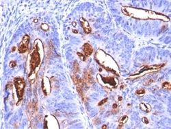
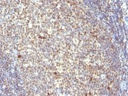
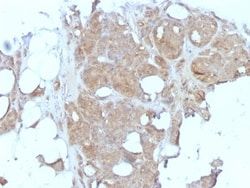
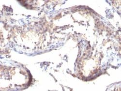
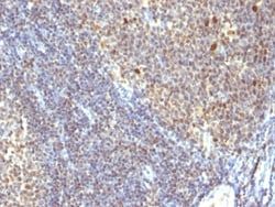
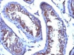
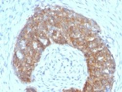
Testosterone Antibody (4E1G2), Novus Biologicals™
Mouse Monoclonal Antibody
Manufacturer: Fischer Scientific
The price for this product is unavailable. Please request a quote
Antigen
Testosterone
Concentration
0.2 mg/mL
Applications
Immunohistochemistry (Paraffin), Radioimmune Assays (RIA), SDS-Page
Conjugate
Unconjugated
Host Species
Mouse
Target Species
Human, All species
Immunogen
Testosterone 3 CMO conjugated to BSA
Primary or Secondary
Primary
Content And Storage
Store at 4C.
Clone
4E1G2
Dilution
Immunohistochemistry-Paraffin 1:10-1:500, Radioimmunoassay, SDS-Page
Classification
Monoclonal
Form
Purified
Regulatory Status
RUO
Formulation
10mM PBS and 0.05% BSA with 0.05% Sodium Azide
Isotype
IgG1 κ
Purification Method
Protein A or G purified
Test Specificity
This MAb is highly specific to testosterone. Its affinity constant for testosterone is ∼1010M-1. In competitive binding immunoassay, it reacts with testosterone 100%, 11-beta-hydroxy testosterone 3.3%, 17-alpha-methyl testosterone
Related Products

Description
- Ensure accurate, reproducible results in ELISA, Radioimmunoassay Testosterone Monoclonal specifically detects Testosterone in Non-species specific samples
- It is validated for ELISA, Immunohistochemistry, Immunohistochemistry-Paraffin, Radioimmunoassay.
Compare Similar Items
Show Difference
Antigen: Testosterone
Concentration: 0.2 mg/mL
Applications: Immunohistochemistry (Paraffin), Radioimmune Assays (RIA), SDS-Page
Conjugate: Unconjugated
Host Species: Mouse
Target Species: Human, All species
Immunogen: Testosterone 3 CMO conjugated to BSA
Primary or Secondary: Primary
Content And Storage: Store at 4C.
Clone: 4E1G2
Dilution: Immunohistochemistry-Paraffin 1:10-1:500, Radioimmunoassay, SDS-Page
Classification: Monoclonal
Form: Purified
Regulatory Status: RUO
Formulation: 10mM PBS and 0.05% BSA with 0.05% Sodium Azide
Isotype: IgG1 κ
Purification Method: Protein A or G purified
Test Specificity: This MAb is highly specific to testosterone. Its affinity constant for testosterone is ∼1010M-1. In competitive binding immunoassay, it reacts with testosterone 100%, 11-beta-hydroxy testosterone 3.3%, 17-alpha-methyl testosterone
Antigen:
Testosterone
Concentration:
0.2 mg/mL
Applications:
Immunohistochemistry (Paraffin), Radioimmune Assays (RIA), SDS-Page
Conjugate:
Unconjugated
Host Species:
Mouse
Target Species:
Human, All species
Immunogen:
Testosterone 3 CMO conjugated to BSA
Primary or Secondary:
Primary
Content And Storage:
Store at 4C.
Clone:
4E1G2
Dilution:
Immunohistochemistry-Paraffin 1:10-1:500, Radioimmunoassay, SDS-Page
Classification:
Monoclonal
Form:
Purified
Regulatory Status:
RUO
Formulation:
10mM PBS and 0.05% BSA with 0.05% Sodium Azide
Isotype:
IgG1 κ
Purification Method:
Protein A or G purified
Test Specificity:
This MAb is highly specific to testosterone. Its affinity constant for testosterone is ∼1010M-1. In competitive binding immunoassay, it reacts with testosterone 100%, 11-beta-hydroxy testosterone 3.3%, 17-alpha-methyl testosterone
Antigen: TFF1/pS2
Concentration: 0.2mg/mL
Applications: Flow Cytometry, Immunohistochemistry (Paraffin), Immunofluorescence
Conjugate: Unconjugated
Host Species: Mouse
Target Species: Human, Cynomolgus Monkey
Immunogen: Synthetic peptide of 28 amino acid residues corresponding to CFDDTVRGVPWCFYPNTIDVPPEEECEF (aa57-84) from the C-terminus of human pS2.
Primary or Secondary: Primary
Content And Storage: Store at 4C.
Clone: SPM313
Dilution: Flow Cytometry 0.5 - 1 ug/million cells in 0.1 ml, Immunohistochemistry-Paraffin 0.5 - 1.0 ug/ml, Immunofluorescence 0.5 - 1.0 ug/ml
Classification: Monoclonal
Form: Purified
Regulatory Status: RUO
Formulation: 10mM PBS and 0.05% BSA with 0.05% Sodium Azide
Isotype: IgG1 κ
Purification Method: Protein A or G purified
Test Specificity: It recognizes a polypeptide of 6.5kDa, identified as pS2 estrogen-regulated protein. Its epitope is localized between aa57-84 of human pS2 protein. pS2 is a trefoil peptide. Trefoil peptides are protease resistant molecules secreted throughout the gut that play a role in mucosal healing. These peptides contain three intra-chain disulfide bonds, forming the trefoil motif, or P-domain. pS2 is known to form dimers and this dimerization is thought to play a role in its protective and healing properties. About 60% of breast carcinomas are positive for pS2. Staining is cytoplasmic, often with localization to the Golgi apparatus. pS2 is shown to be localized in normal stomach mucosa, gastric fluid, goblet cells in the colon and small intestine, and in ulcerations of the gastrointestinal tract. Several studies have shown that pS2 is primarily expressed in estrogen receptor-positive breast tumors and it may define a subset of estrogen-dependent tumors that displays an increased likelihood of re
Antigen:
TFF1/pS2
Concentration:
0.2mg/mL
Applications:
Flow Cytometry, Immunohistochemistry (Paraffin), Immunofluorescence
Conjugate:
Unconjugated
Host Species:
Mouse
Target Species:
Human, Cynomolgus Monkey
Immunogen:
Synthetic peptide of 28 amino acid residues corresponding to CFDDTVRGVPWCFYPNTIDVPPEEECEF (aa57-84) from the C-terminus of human pS2.
Primary or Secondary:
Primary
Content And Storage:
Store at 4C.
Clone:
SPM313
Dilution:
Flow Cytometry 0.5 - 1 ug/million cells in 0.1 ml, Immunohistochemistry-Paraffin 0.5 - 1.0 ug/ml, Immunofluorescence 0.5 - 1.0 ug/ml
Classification:
Monoclonal
Form:
Purified
Regulatory Status:
RUO
Formulation:
10mM PBS and 0.05% BSA with 0.05% Sodium Azide
Isotype:
IgG1 κ
Purification Method:
Protein A or G purified
Test Specificity:
It recognizes a polypeptide of 6.5kDa, identified as pS2 estrogen-regulated protein. Its epitope is localized between aa57-84 of human pS2 protein. pS2 is a trefoil peptide. Trefoil peptides are protease resistant molecules secreted throughout the gut that play a role in mucosal healing. These peptides contain three intra-chain disulfide bonds, forming the trefoil motif, or P-domain. pS2 is known to form dimers and this dimerization is thought to play a role in its protective and healing properties. About 60% of breast carcinomas are positive for pS2. Staining is cytoplasmic, often with localization to the Golgi apparatus. pS2 is shown to be localized in normal stomach mucosa, gastric fluid, goblet cells in the colon and small intestine, and in ulcerations of the gastrointestinal tract. Several studies have shown that pS2 is primarily expressed in estrogen receptor-positive breast tumors and it may define a subset of estrogen-dependent tumors that displays an increased likelihood of re
Antigen: TfR (Transferrin R)
Concentration: 0.2 mg/mL
Applications: Flow Cytometry, Immunofluorescence
Conjugate: Unconjugated
Host Species: Mouse
Target Species: Human
Immunogen: Recombinant human TFRC protein
Primary or Secondary: Primary
Content And Storage: Store at 4C.
Clone: TFRC/1059
Dilution: Flow Cytometry 0.5 - 1 ug/million cells in 0.1 ml, Immunofluorescence 1 - 2 ug/ml
Classification: Monoclonal
Form: Purified
Regulatory Status: RUO
Formulation: 10mM PBS and 0.05% BSA with 0.05% Sodium Azide
Isotype: IgG1 κ
Purification Method: Protein A or G purified
Test Specificity: It recognizes a ∼90-95kDa protein which is identified as cell surface transferrin receptor (CD71), a disulfide-bonded homodimeric glycoprotein of 180-190kDa. This MAb is highly specific to CD71 and shows no cross-reaction with other related proteins. Its epitope is localized in the extracellular domain of CD71. Ligand for transferrin receptor is the serum iron transport protein, transferrin. This receptor is broadly distributed in carcinomas, sarcomas, leukemias, and lymphomas. CD71/Transferrin receptor has been reported to be associated with cell proliferation in both normal and neoplastic tissues and useful in predicting clinical behavior or response to therapy in a number of malignancies including breast cancer.
Antigen:
TfR (Transferrin R)
Concentration:
0.2 mg/mL
Applications:
Flow Cytometry, Immunofluorescence
Conjugate:
Unconjugated
Host Species:
Mouse
Target Species:
Human
Immunogen:
Recombinant human TFRC protein
Primary or Secondary:
Primary
Content And Storage:
Store at 4C.
Clone:
TFRC/1059
Dilution:
Flow Cytometry 0.5 - 1 ug/million cells in 0.1 ml, Immunofluorescence 1 - 2 ug/ml
Classification:
Monoclonal
Form:
Purified
Regulatory Status:
RUO
Formulation:
10mM PBS and 0.05% BSA with 0.05% Sodium Azide
Isotype:
IgG1 κ
Purification Method:
Protein A or G purified
Test Specificity:
It recognizes a ∼90-95kDa protein which is identified as cell surface transferrin receptor (CD71), a disulfide-bonded homodimeric glycoprotein of 180-190kDa. This MAb is highly specific to CD71 and shows no cross-reaction with other related proteins. Its epitope is localized in the extracellular domain of CD71. Ligand for transferrin receptor is the serum iron transport protein, transferrin. This receptor is broadly distributed in carcinomas, sarcomas, leukemias, and lymphomas. CD71/Transferrin receptor has been reported to be associated with cell proliferation in both normal and neoplastic tissues and useful in predicting clinical behavior or response to therapy in a number of malignancies including breast cancer.

Antigen: TfR (Transferrin R)
Concentration: 0.2 mg/mL
Applications: Western Blot, Flow Cytometry, Immunofluorescence
Conjugate: Unconjugated
Host Species: Mouse
Target Species: Human
Immunogen: Recombinant human TFRC protein
Primary or Secondary: Primary
Content And Storage: Store at 4C.
Clone: TFRC/1149
Dilution: Western Blot 0.5 - 1.0 ug/ml, Flow Cytometry 0.5 - 1 ug/million cells in 0.1 ml, Immunofluorescence 1 - 2 ug/ml
Classification: Monoclonal
Form: Purified
Regulatory Status: RUO
Formulation: 10mM PBS and 0.05% BSA with 0.05% Sodium Azide
Isotype: IgG1 κ
Purification Method: Protein A or G purified
Test Specificity: It recognizes a ∼90-95kDa protein which is identified as cell surface transferrin receptor (CD71), a disulfide-bonded homodimeric glycoprotein of 180-190kDa. This MAb is highly specific to CD71 and shows no cross-reaction with other related proteins. Ligand for transferrin receptor is the serum iron transport protein, transferrin. This receptor is broadly distributed in carcinomas, sarcomas, leukemias, and lymphomas. CD71/Transferrin receptor has been reported to be associated with cell proliferation in both normal and neoplastic tissues and useful in predicting clinical behavior or response to therapy in a number of malignancies including breast cancer.
Antigen:
TfR (Transferrin R)
Concentration:
0.2 mg/mL
Applications:
Western Blot, Flow Cytometry, Immunofluorescence
Conjugate:
Unconjugated
Host Species:
Mouse
Target Species:
Human
Immunogen:
Recombinant human TFRC protein
Primary or Secondary:
Primary
Content And Storage:
Store at 4C.
Clone:
TFRC/1149
Dilution:
Western Blot 0.5 - 1.0 ug/ml, Flow Cytometry 0.5 - 1 ug/million cells in 0.1 ml, Immunofluorescence 1 - 2 ug/ml
Classification:
Monoclonal
Form:
Purified
Regulatory Status:
RUO
Formulation:
10mM PBS and 0.05% BSA with 0.05% Sodium Azide
Isotype:
IgG1 κ
Purification Method:
Protein A or G purified
Test Specificity:
It recognizes a ∼90-95kDa protein which is identified as cell surface transferrin receptor (CD71), a disulfide-bonded homodimeric glycoprotein of 180-190kDa. This MAb is highly specific to CD71 and shows no cross-reaction with other related proteins. Ligand for transferrin receptor is the serum iron transport protein, transferrin. This receptor is broadly distributed in carcinomas, sarcomas, leukemias, and lymphomas. CD71/Transferrin receptor has been reported to be associated with cell proliferation in both normal and neoplastic tissues and useful in predicting clinical behavior or response to therapy in a number of malignancies including breast cancer.