Endorepellin/Perlecan/Heparan Sulfate Proteoglycan Antibody (A7L6), Novus Biologicals™
Rat Monoclonal Antibody has been used in 1 publication
Manufacturer: Fischer Scientific
The price for this product is unavailable. Please request a quote
Antigen
Endorepellin/Perlecan/Heparan Sulfate Proteoglycan
Concentration
0.2mg/mL
Applications
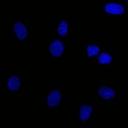
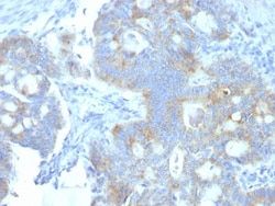
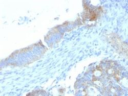
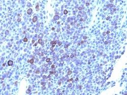
Flow Cytometry, Immunohistochemistry (Paraffin), SDS-Page, Immunofluorescence
Conjugate
Unconjugated
Host Species
Rat
Target Species
Human, Mouse, Porcine, Fish, Monkey
Gene Accession No.
P98160
Gene ID (Entrez)
3339
Immunogen
Murine EHS laminin preparation
Primary or Secondary
Primary
Content And Storage
Store at 4C.
Clone
A7L6
Dilution
Flow Cytometry 0.5 - 1 ug/million cells in 0.1 ml, Immunohistochemistry-Paraffin 1 - 2 ug/ml, SDS-Page, Immunofluorescence 0.5 - 1.0 ug/ml
Classification
Monoclonal
Form
Purified
Regulatory Status
RUO
Formulation
10mM PBS and 0.05% BSA with 0.05% Sodium Azide
Gene Alias
basement membrane-specific heparan sulfate proteoglycan core protein, endorepellin (domain V region), heparan sulfate proteoglycan 2, HSPG, perlecan, perlecan proteoglycan, PLCSchwartz-Jampel syndrome 1 (chondrodystrophic myotonia), PRCAN, SJA, SJS, SJS1
Gene Symbols
HSPG2
Isotype
IgG2a κ
Purification Method
Protein A or G purified
Test Specificity
This MAb specifically precipitates heterogeneous material of high MW, identified as perlecan, a major heparan-sulfate proteoglycan (HSPG) within all basement membranes and cell surfaces. It does not cross-react with laminin, fibronectin, or dermatran sulfate proteoglycan. Because of perlecan s strategic location and ability to store and protect growth factors, it has been strongly implicated in the control of tumor cell growth and metastatic behavior. Perlecan possesses angiogenic and growth-promoting attributes primarily by acting as a co-receptor for basic fibroblast growth factor (FGF-2). Suppression of perlecan causes substantial inhibition of neoplastic growth and neovascularization. Thus, perlecan is a potent inducer of neoplasm growth and angiogenesis in vivo and therapeutic interventions targeting this key modulator of tumor progression may improve neoplastic treatment.
Related Products

Description
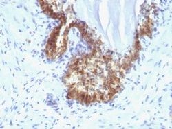
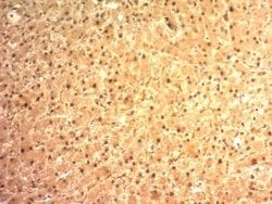
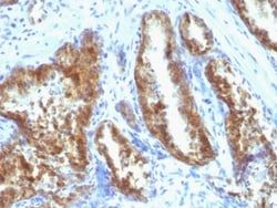
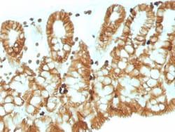
- Ensure accurate, reproducible results in Flow Cytometry, Immunohistochemistry (Paraffin), Immunofluorescence Endorepellin/Perlecan/Heparan Sulfate Proteoglycan Monoclonal specifically detects Endorepellin/Perlecan/Heparan Sulfate Proteoglycan in Human, Mouse, Porcine, Bovine, Fish, Monkey samples
- It is validated for Flow Cytometry, Immunohistochemistry, Immunocytochemistry/Immunofluorescence, Immunohistochemistry-Paraffin, Immunohistochemistry-Frozen, Immunofluorescence.
Compare Similar Items
Show Difference
Antigen: Endorepellin/Perlecan/Heparan Sulfate Proteoglycan
Concentration: 0.2mg/mL
Applications: Flow Cytometry, Immunohistochemistry (Paraffin), SDS-Page, Immunofluorescence
Conjugate: Unconjugated
Host Species: Rat
Target Species: Human, Mouse, Porcine, Fish, Monkey
Gene Accession No.: P98160
Gene ID (Entrez): 3339
Immunogen: Murine EHS laminin preparation
Primary or Secondary: Primary
Content And Storage: Store at 4C.
Clone: A7L6
Dilution: Flow Cytometry 0.5 - 1 ug/million cells in 0.1 ml, Immunohistochemistry-Paraffin 1 - 2 ug/ml, SDS-Page, Immunofluorescence 0.5 - 1.0 ug/ml
Classification: Monoclonal
Form: Purified
Regulatory Status: RUO
Formulation: 10mM PBS and 0.05% BSA with 0.05% Sodium Azide
Gene Alias: basement membrane-specific heparan sulfate proteoglycan core protein, endorepellin (domain V region), heparan sulfate proteoglycan 2, HSPG, perlecan, perlecan proteoglycan, PLCSchwartz-Jampel syndrome 1 (chondrodystrophic myotonia), PRCAN, SJA, SJS, SJS1
Gene Symbols: HSPG2
Isotype: IgG2a κ
Purification Method: Protein A or G purified
Test Specificity: This MAb specifically precipitates heterogeneous material of high MW, identified as perlecan, a major heparan-sulfate proteoglycan (HSPG) within all basement membranes and cell surfaces. It does not cross-react with laminin, fibronectin, or dermatran sulfate proteoglycan. Because of perlecan s strategic location and ability to store and protect growth factors, it has been strongly implicated in the control of tumor cell growth and metastatic behavior. Perlecan possesses angiogenic and growth-promoting attributes primarily by acting as a co-receptor for basic fibroblast growth factor (FGF-2). Suppression of perlecan causes substantial inhibition of neoplastic growth and neovascularization. Thus, perlecan is a potent inducer of neoplasm growth and angiogenesis in vivo and therapeutic interventions targeting this key modulator of tumor progression may improve neoplastic treatment.
Antigen: Endorepellin/Perlecan/Heparan Sulfate Proteoglycan
Concentration: 0.2mg/mL
Applications: Flow Cytometry, Immunohistochemistry (Paraffin), Immunofluorescence
Conjugate: Unconjugated
Host Species: Rat
Target Species: Human, Mouse, Porcine, Fish, Monkey, Primate
Gene Accession No.: __
Gene ID (Entrez): 3339
Immunogen: Murine EHS laminin preparation
Primary or Secondary: Primary
Content And Storage: Store at 4C.
Clone: SPM255
Dilution: Flow Cytometry 0.5 - 1 ug/million cells in 0.1 ml, Immunohistochemistry-Paraffin 1 - 2 ug/ml, Immunofluorescence 0.5 - 1.0 ug/ml
Classification: Monoclonal
Form: Purified
Regulatory Status: RUO
Formulation: 10mM PBS and 0.05% BSA with 0.05% Sodium Azide
Gene Alias: basement membrane-specific heparan sulfate proteoglycan core protein, endorepellin (domain V region), heparan sulfate proteoglycan 2, HSPG, perlecan, perlecan proteoglycan, PLCSchwartz-Jampel syndrome 1 (chondrodystrophic myotonia), PRCAN, SJA, SJS, SJS1
Gene Symbols: HSPG2
Isotype: IgG2a κ
Purification Method: Protein A or G purified
Test Specificity: This MAb specifically precipitates heterogeneous material of high MW, identified as perlecan, a major heparan-sulfate proteoglycan (HSPG) within all basement membranes and cell surfaces. It does not cross-react with laminin, fibronectin, or dermatran sulfate proteoglycan. Because of perlecan s strategic location and ability to store and protect growth factors, it has been strongly implicated in the control of tumor cell growth and metastatic behavior. Perlecan possesses angiogenic and growth-promoting attributes primarily by acting as a co-receptor for basic fibroblast growth factor (FGF-2). Suppression of perlecan causes substantial inhibition of neoplastic growth and neovascularization. Thus, perlecan is a potent inducer of neoplasm growth and angiogenesis in vivo and therapeutic interventions targeting this key modulator of tumor progression may improve neoplastic treatment
Antigen: EpCAM/TROP1
Concentration: 0.2mg/mL
Applications: Western Blot, Flow Cytometry, Immunohistochemistry (Paraffin), Immunofluorescence
Conjugate: Unconjugated
Host Species: Mouse
Target Species: Human, Mouse, Rat
Gene Accession No.: P16422
Gene ID (Entrez): 4072
Immunogen: A recombinant human EpCAM fragment from the cytoplasmic domain (around aa 280-350) (exact sequence is proprietary)
Primary or Secondary: Primary
Content And Storage: Store at 4C.
Clone: EGP40/1110
Dilution: Western Blot 0.5 - 1.0 ug/ml, Flow Cytometry 0.5 - 1 ug/million cells in 0.1 ml, Immunohistochemistry-Paraffin 0.5 - 1.0 ug/ml, Immunofluorescence 1 - 2 ug/ml
Classification: Monoclonal
Form: Purified
Regulatory Status: RUO
Formulation: 10mM PBS and 0.05% BSA with 0.05% Sodium Azide
Gene Alias: 17-1A, 323/A3, ACSTD1, antigen identified by monoclonal AUA1, CD326 antigen, Cell surface glycoprotein Trop-1, chromosome 4, surface marker (35kD glycoprotein), DIAR5, EGP, EGP-2, EGP314, EGP40, EpCAM, epithelial cell adhesion molecule, Epithelial cell surface antigen, Epithelial glycoprotein, Epithelial glycoprotein 314, ESA, GA733-2EGP34, hEGP314, HNPCC8, KS 1/4 antigen, KS1/4, KSAHEA125, M1S2, M4S1Ly74, Major gastrointestinal tumor-associated protein GA733-2, MIC18MH99, MOC31, TACST-1, TACSTD1, TROP1CD326, Tumor-associated calcium signal transducer 1CO-17A
Gene Symbols: EPCAM
Isotype: IgG2b κ
Purification Method: Protein A or G purified
Test Specificity: EGP40 is a 40-43kDa transmembrane epithelial glycoprotein, also identified as epithelial specific antigen (ESA), or epithelial cellular adhesion molecule (Ep-CAM). It is expressed on baso-lateral cell surface in most simple epithelia and a vast majority of carcinomas. This antibody has been used to distinguish adenocarcinoma from pleural mesothelioma and hepatocellular carcinoma. This antibody is also useful in distinguishing serous carcinomas of the ovary from mesothelioma.