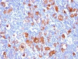
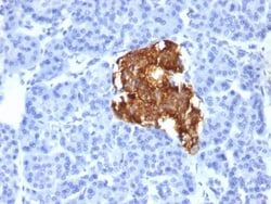
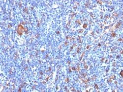
Fas/TNFRSF6/CD95 Antibody (B-R18), Novus Biologicals™
Mouse Monoclonal Antibody
Manufacturer: Fischer Scientific
The price for this product is unavailable. Please request a quote
Antigen
Fas/TNFRSF6/CD95
Concentration
0.2 mg/mL
Applications
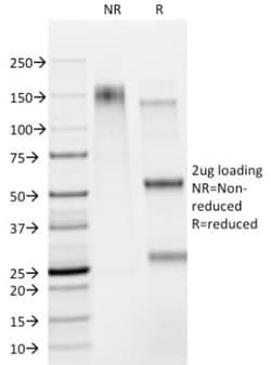
Flow Cytometry, Immunohistochemistry (Frozen), SDS-Page, Immunofluorescence
Conjugate
Unconjugated
Host Species
Mouse
Research Discipline
Apoptosis, Cancer, Cell Biology, Death Receptor Signaling Pathway, Diabetes Research, Inhibitors Activators and Regulators
Formulation
10mM PBS and 0.05% BSA with 0.05% Sodium Azide
Gene Alias
Apo-1 antigen, apoptosis antigen 1, Apoptosis-mediating surface antigen FAS, APT1FASTM, CD95 antigen, CD95ALPS1A, Fas (TNF receptor superfamily, member 6), Fas AMA, Fas antigen, FAS1, FASLG receptor, TNFRSF6member 6, tumor necrosis factor receptor superfamily member 6
Gene Symbols
FAS
Isotype
IgG1 κ
Purification Method
Protein A or G purified
Test Specificity
MAb B-R18 specifically recognizes CD95, also known as Fas, a transmembrane glycoprotein with a MW of 40-45kDa, containing 8kDa of N-glycosidic-linked polysaccharide. It is a receptor for TNFSF6/FASLG, a member of the nerve growth factor receptor/tumor necrosis factor superfamily, mediating receptor-triggered apoptosis. The adapter molecule FADD recruits caspase-8 to the activated receptor. The resulting death-inducing signaling complex (DISC) performs caspase-8 proteolytic activation, which initiates the subsequent cascade of caspases (aspartate-specific cysteine proteases) mediating apoptosis. FAS-mediated apoptosis may have a role in the induction of peripheral tolerance, in the antigen-stimulated suicide of mature T-cells, or both. The secreted isoforms 2 to 6 block apoptosis (in vitro). CD95 antigen is expressed on the surface of various cell types, preferentially on the CD45RAlow CD45ROhigh subset of memory T lymphocytes.
Clone
B-R18
Dilution
Flow Cytometry 0.5 - 1 ug/million cells in 0.1 ml, Immunohistochemistry-Frozen 0.5 - 1.0 ug/ml, SDS-Page, Immunofluorescence 0.5 - 1.0 ug/ml
Classification
Monoclonal
Form
Purified
Regulatory Status
RUO
Target Species
Human
Gene Accession No.
P25445
Gene ID (Entrez)
355
Immunogen
Recombinant human CD95 protein
Primary or Secondary
Primary
Content And Storage
Store at 4C.
Related Products


Description
- Ensure accurate, reproducible results in Flow Cytometry, Immunohistochemistry (Frozen), Immunofluorescence Fas Receptor/TNFRSF6/CD95 Monoclonal specifically detects Fas Receptor/TNFRSF6/CD95 in Human samples
- It is validated for Flow Cytometry, Immunohistochemistry, Immunocytochemistry/Immunofluorescence, Immunohistochemistry-Frozen, Functional, Immunofluorescence.
Compare Similar Items
Show Difference
Antigen: Fas/TNFRSF6/CD95
Concentration: 0.2 mg/mL
Applications: Flow Cytometry, Immunohistochemistry (Frozen), SDS-Page, Immunofluorescence
Conjugate: Unconjugated
Host Species: Mouse
Research Discipline: Apoptosis, Cancer, Cell Biology, Death Receptor Signaling Pathway, Diabetes Research, Inhibitors Activators and Regulators
Formulation: 10mM PBS and 0.05% BSA with 0.05% Sodium Azide
Gene Alias: Apo-1 antigen, apoptosis antigen 1, Apoptosis-mediating surface antigen FAS, APT1FASTM, CD95 antigen, CD95ALPS1A, Fas (TNF receptor superfamily, member 6), Fas AMA, Fas antigen, FAS1, FASLG receptor, TNFRSF6member 6, tumor necrosis factor receptor superfamily member 6
Gene Symbols: FAS
Isotype: IgG1 κ
Purification Method: Protein A or G purified
Test Specificity: MAb B-R18 specifically recognizes CD95, also known as Fas, a transmembrane glycoprotein with a MW of 40-45kDa, containing 8kDa of N-glycosidic-linked polysaccharide. It is a receptor for TNFSF6/FASLG, a member of the nerve growth factor receptor/tumor necrosis factor superfamily, mediating receptor-triggered apoptosis. The adapter molecule FADD recruits caspase-8 to the activated receptor. The resulting death-inducing signaling complex (DISC) performs caspase-8 proteolytic activation, which initiates the subsequent cascade of caspases (aspartate-specific cysteine proteases) mediating apoptosis. FAS-mediated apoptosis may have a role in the induction of peripheral tolerance, in the antigen-stimulated suicide of mature T-cells, or both. The secreted isoforms 2 to 6 block apoptosis (in vitro). CD95 antigen is expressed on the surface of various cell types, preferentially on the CD45RAlow CD45ROhigh subset of memory T lymphocytes.
Clone: B-R18
Dilution: Flow Cytometry 0.5 - 1 ug/million cells in 0.1 ml, Immunohistochemistry-Frozen 0.5 - 1.0 ug/ml, SDS-Page, Immunofluorescence 0.5 - 1.0 ug/ml
Classification: Monoclonal
Form: Purified
Regulatory Status: RUO
Target Species: Human
Gene Accession No.: P25445
Gene ID (Entrez): 355
Immunogen: Recombinant human CD95 protein
Primary or Secondary: Primary
Content And Storage: Store at 4C.
Antigen: Fascin
Concentration: 0.2mg/mL
Applications: Western Blot, Flow Cytometry, Immunohistochemistry (Paraffin), SDS-Page, Immunofluorescence
Conjugate: Unconjugated
Host Species: Mouse
Research Discipline: Cytoskeleton Markers
Formulation: 10mM PBS and 0.05% BSA with 0.05% Sodium Azide
Gene Alias: FAN1,55 kDa actin-bundling protein, fascin homolog 1, actin-bundling protein (Strongylocentrotus purpuratus), FLJ38511, HSN, p55actin bundling protein, singed (Drosophila)-like (sea urchin fascin homolog like), singed-like (fascin homolog, sea urchin), Singed-like protein, SNLfascin
Gene Symbols: FSCN1
Isotype: IgG2a κ
Purification Method: Protein A or G purified
Test Specificity: Recognizes a protein of 55kDa, which is identified as fascin-1. Its actin binding ability is regulated by phosphorylation. Antibody to fascin-1 is a very sensitive marker for Reed-Sternberg cells and variants in nodular sclerosis, mixed cellularity, and lymphocyte depletion Hodgkin's disease. It is uniformly negative in lymphoid cells, plasma cells, and myeloid cells. Fascin-1 is also expressed in dendritic cells. This marker may be helpful to distinguish between Hodgkin lymphoma and non-Hodgkin lymphoma in difficult cases. Also, the lack of expression of fascin-1 in the neoplastic follicles in follicular lymphoma may be helpful in distinguishing these lymphomas from reactive follicular hyperplasia in which the number of follicular dendritic cells is normal or increased. Antibody to fascin-1 has been suggested as a prognostic marker in neuroendocrine neoplasms of the lung as well as in ovarian cancer. Fascin-1 expression may be induced by Epstein-Barr virus (EBV) infection of B cells w
Clone: FSCN1/417
Dilution: Western Blot 0.5 - 1.0 ug/ml, Simple Western 10 ug/ml, Flow Cytometry 0.5 - 1 ug/million cells in 0.1 ml, Immunohistochemistry-Paraffin 0.5 - 1.0 ug/ml, SDS-Page, Immunofluorescence 1 - 2 ug/ml
Classification: Monoclonal
Form: Purified
Regulatory Status: RUO
Target Species: Human
Gene Accession No.: Q16658
Gene ID (Entrez): 6624
Immunogen: Full length recombinant human FSCN1 protein
Primary or Secondary: Primary
Content And Storage: Store at 4C.