CD43/Sialophorin Antibody (84-3C1), Novus Biologicals™
Mouse Monoclonal Antibody
Manufacturer: Fischer Scientific
The price for this product is unavailable. Please request a quote
Antigen
CD43/Sialophorin
Concentration
0.2mg/mL
Applications
Flow Cytometry, Immunohistochemistry (Paraffin), Immunofluorescence
Conjugate
Unconjugated
Host Species
Mouse
Research Discipline
Immunology
Formulation
10mM PBS and 0.05% BSA with 0.05% Sodium Azide
Gene ID (Entrez)
6693
Immunogen
Stimulated human leukocytes
Primary or Secondary
Primary
Content And Storage
Store at 4C.
Clone
84-3C1
Dilution
Flow Cytometry 0.5 - 1 ug/million cells in 0.1 ml, Immunohistochemistry-Paraffin 0.5 - 1.0 ug/ml, Immunofluorescence 1 - 2 ug/ml
Classification
Monoclonal
Form
Purified
Regulatory Status
RUO
Target Species
Human
Gene Alias
CD43 antigen, CD43), Galactoglycoprotein, GALGP, Leukocyte sialoglycoprotein, Sialophorin, sialophorin (gpL115, leukosialin, CD43)
Gene Symbols
SPN
Isotype
IgG1 κ
Purification Method
Protein A or G purified
Test Specificity
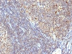
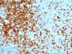
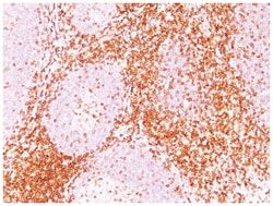
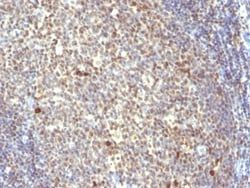
It recognizes a cell surface glycoprotein of 95/115/135kDa (depending upon the extent of glycosylation), identified as CD43 (Workshop III). 70-90% of T-cell lymphomas and from 22-37% of B-cell lymphomas express CD43. No reactivity has been observed with reactive B-cells. So a B-lineage population that co-expresses CD43 is highly likely to be a malignant lymphoma, especially a low-grade lymphoma, rather than a reactive B-cell population. When CD43 antibody is used in combination with anti-CD20, effective immunophenotyping of the lymphomas in formalin-fixed tissues can be obtained. Co-staining of a lymphoid infiltrate with anti-CD20 and anti-CD43 argues against a reactive process and favors a diagnosis of lymphoma.
Related Products


Description
- Ensure accurate, reproducible results in Flow Cytometry, Immunohistochemistry (Paraffin), Immunofluorescence CD43/Sialophorin Monoclonal specifically detects CD43/Sialophorin in Human samples
- It is validated for Western Blot, Flow Cytometry, Immunohistochemistry, Immunocytochemistry/Immunofluorescence, Immunohistochemistry-Paraffin.
Compare Similar Items
Show Difference
Antigen: CD43/Sialophorin
Concentration: 0.2mg/mL
Applications: Flow Cytometry, Immunohistochemistry (Paraffin), Immunofluorescence
Conjugate: Unconjugated
Host Species: Mouse
Research Discipline: Immunology
Formulation: 10mM PBS and 0.05% BSA with 0.05% Sodium Azide
Gene ID (Entrez): 6693
Immunogen: Stimulated human leukocytes
Primary or Secondary: Primary
Content And Storage: Store at 4C.
Clone: 84-3C1
Dilution: Flow Cytometry 0.5 - 1 ug/million cells in 0.1 ml, Immunohistochemistry-Paraffin 0.5 - 1.0 ug/ml, Immunofluorescence 1 - 2 ug/ml
Classification: Monoclonal
Form: Purified
Regulatory Status: RUO
Target Species: Human
Gene Alias: CD43 antigen, CD43), Galactoglycoprotein, GALGP, Leukocyte sialoglycoprotein, Sialophorin, sialophorin (gpL115, leukosialin, CD43)
Gene Symbols: SPN
Isotype: IgG1 κ
Purification Method: Protein A or G purified
Test Specificity: It recognizes a cell surface glycoprotein of 95/115/135kDa (depending upon the extent of glycosylation), identified as CD43 (Workshop III). 70-90% of T-cell lymphomas and from 22-37% of B-cell lymphomas express CD43. No reactivity has been observed with reactive B-cells. So a B-lineage population that co-expresses CD43 is highly likely to be a malignant lymphoma, especially a low-grade lymphoma, rather than a reactive B-cell population. When CD43 antibody is used in combination with anti-CD20, effective immunophenotyping of the lymphomas in formalin-fixed tissues can be obtained. Co-staining of a lymphoid infiltrate with anti-CD20 and anti-CD43 argues against a reactive process and favors a diagnosis of lymphoma.
Antigen: CA19-9/Sialyl Lewisa
Concentration: 0.2mg/mL
Applications: Flow Cytometry, Immunohistochemistry (Paraffin), Immunofluorescence
Conjugate: Unconjugated
Host Species: Mouse
Research Discipline: __
Formulation: 10mM PBS and 0.05% BSA with 0.05% Sodium Azide
Gene ID (Entrez): __
Immunogen: Precipitin lines obtained after immuno-diffusion using MAb 116-NS-19-9 and mucins isolated from an ovarian cyst of a Lewis A+B- patient (0Le).
Primary or Secondary: Primary
Content And Storage: Store at 4C.
Clone: SPM588
Dilution: Flow Cytometry 0.5 - 1 ug/million cells in 0.1 ml, Immunohistochemistry-Paraffin 0.5 - 1.0 ug/ml, Immunofluorescence 0.5 - 1.0 ug/ml
Classification: Monoclonal
Form: Purified
Regulatory Status: RUO
Target Species: Human
Gene Alias: __
Gene Symbols: __
Isotype: IgM κ
Purification Method: Protein A or G purified
Test Specificity: __
Antigen: IPO-38
Concentration: 0.2mg/mL
Applications: Flow Cytometry, Immunohistochemistry (Paraffin), Immunofluorescence
Conjugate: Unconjugated
Host Species: Mouse
Research Discipline: __
Formulation: 10mM PBS and 0.05% BSA with 0.05% Sodium Azide
Gene ID (Entrez): __
Immunogen: Spleen cells of a patient with hairy cell leukemia
Primary or Secondary: Primary
Content And Storage: Store at 4C.
Clone: IPO-38
Dilution: Flow Cytometry 5 - 10 ul/million cells in 0.1ml, Immunohistochemistry-Paraffin 1:25 - 1:50, Immunofluorescence 1:25 - 1:50
Classification: Monoclonal
Form: Purified
Regulatory Status: RUO
Target Species: Human, Mouse, Rat
Gene Alias: __
Gene Symbols: __
Isotype: IgM κ
Purification Method: Protein A or G purified
Test Specificity: Recognizes a protein of 14-16kDa, which is a novel nuclear antigen of proliferating cells. IPO-38 antigen is present in the nuclei of proliferating cells such as Hodgkin s disease and non-Hodgkin s lymphomas, different forms of leukemias, breast and colorectal carcinomas, and PHA-stimulated lymphocytes. It is not expressed in the cells of non-stimulated lymphocytes and granulocytes. IPO-38 may be a useful marker of cell proliferation during monitoring of tumor progression.