CD99 Antibody (SPM586), Novus Biologicals™
Mouse Monoclonal Antibody
Manufacturer: Fischer Scientific
The price for this product is unavailable. Please request a quote
Antigen
CD99
Concentration
0.2mg/mL
Applications
Flow Cytometry, Immunohistochemistry (Paraffin), Immunofluorescence, CyTOF
Conjugate
Unconjugated
Host Species
Mouse
Research Discipline
Immunology
Formulation
10mM PBS with 0.05% Sodium Azide
Gene ID (Entrez)
4267
Immunogen
Purified E-rosette forming cells from human peripheral blood lymphocytes
Primary or Secondary
Primary
Content And Storage
Store at 4C.
Clone
SPM586
Dilution
Flow Cytometry 5 - 10ul/million cells in 0.1ml, Immunohistochemistry-Paraffin 1 - 2ug/ml, Immunofluorescence 0.5 - 1.0ug/ml, CyTOF-ready
Classification
Monoclonal
Form
Purified
Regulatory Status
RUO
Target Species
Human, Rat
Gene Alias
12E7, antigen identified by monoclonal antibodies 12E7, F21 and O13, CD99 antigenY homolog, CD99 molecule, E2 antigen, HBA71, MIC2 (monoclonal 12E7), MIC2Y, MSK5X, Protein MIC2, surface antigen MIC2, T-cell surface glycoprotein E2
Gene Symbols
CD99
Isotype
IgM κ
Purification Method
Protein A or G purified
Test Specificity
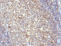
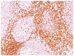
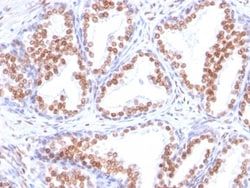
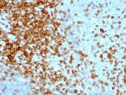
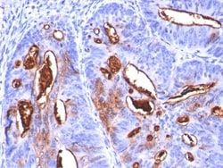
Recognizes a sialoglycoprotein of 27-32kDa, identified as CD99, or MIC2 gene product, or E2 antigen. This antigen is expressed on the cell membrane of some lymphocytes, cortical thymocytes, and granulosa cells of the ovary. Most pancreatic islet cells, Sertoli cells of the testis, and some endothelial cells express this antigen. Mature granulocytes express very little or no CD99. MIC2 is strongly expressed on Ewing s sarcoma cells and primitive peripheral neuroectodermal tumors. This MAb shows a very similar reactivity to other CD99 MAbs (e.g. O13, 12E7, or HBA-71) and is excellent for immunohistochemical staining of formalin-fixed, paraffin-embedded tissues.
Related Products


Description
- Ensure accurate, reproducible results in Flow Cytometry, Immunohistochemistry (Paraffin), Immunofluorescence CD99 Monoclonal specifically detects CD99 in Human, Rat samples
- It is validated for Flow Cytometry, Immunohistochemistry, Immunocytochemistry/Immunofluorescence, Immunohistochemistry-Paraffin, Immunofluorescence, CyTOF-ready.
Compare Similar Items
Show Difference
Antigen: CD99
Concentration: 0.2mg/mL
Applications: Flow Cytometry, Immunohistochemistry (Paraffin), Immunofluorescence, CyTOF
Conjugate: Unconjugated
Host Species: Mouse
Research Discipline: Immunology
Formulation: 10mM PBS with 0.05% Sodium Azide
Gene ID (Entrez): 4267
Immunogen: Purified E-rosette forming cells from human peripheral blood lymphocytes
Primary or Secondary: Primary
Content And Storage: Store at 4C.
Clone: SPM586
Dilution: Flow Cytometry 5 - 10ul/million cells in 0.1ml, Immunohistochemistry-Paraffin 1 - 2ug/ml, Immunofluorescence 0.5 - 1.0ug/ml, CyTOF-ready
Classification: Monoclonal
Form: Purified
Regulatory Status: RUO
Target Species: Human, Rat
Gene Alias: 12E7, antigen identified by monoclonal antibodies 12E7, F21 and O13, CD99 antigenY homolog, CD99 molecule, E2 antigen, HBA71, MIC2 (monoclonal 12E7), MIC2Y, MSK5X, Protein MIC2, surface antigen MIC2, T-cell surface glycoprotein E2
Gene Symbols: CD99
Isotype: IgM κ
Purification Method: Protein A or G purified
Test Specificity: Recognizes a sialoglycoprotein of 27-32kDa, identified as CD99, or MIC2 gene product, or E2 antigen. This antigen is expressed on the cell membrane of some lymphocytes, cortical thymocytes, and granulosa cells of the ovary. Most pancreatic islet cells, Sertoli cells of the testis, and some endothelial cells express this antigen. Mature granulocytes express very little or no CD99. MIC2 is strongly expressed on Ewing s sarcoma cells and primitive peripheral neuroectodermal tumors. This MAb shows a very similar reactivity to other CD99 MAbs (e.g. O13, 12E7, or HBA-71) and is excellent for immunohistochemical staining of formalin-fixed, paraffin-embedded tissues.
Antigen: CD43/Sialophorin
Concentration: 0.2mg/mL
Applications: Flow Cytometry, Immunohistochemistry (Paraffin), Immunofluorescence
Conjugate: Unconjugated
Host Species: Mouse
Research Discipline: Immunology
Formulation: 10mM PBS and 0.05% BSA with 0.05% Sodium Azide
Gene ID (Entrez): 6693
Immunogen: Stimulated human leukocytes
Primary or Secondary: Primary
Content And Storage: Store at 4C.
Clone: 84-3C1
Dilution: Flow Cytometry 0.5 - 1 ug/million cells in 0.1 ml, Immunohistochemistry-Paraffin 0.5 - 1.0 ug/ml, Immunofluorescence 1 - 2 ug/ml
Classification: Monoclonal
Form: Purified
Regulatory Status: RUO
Target Species: Human
Gene Alias: CD43 antigen, CD43), Galactoglycoprotein, GALGP, Leukocyte sialoglycoprotein, Sialophorin, sialophorin (gpL115, leukosialin, CD43)
Gene Symbols: SPN
Isotype: IgG1 κ
Purification Method: Protein A or G purified
Test Specificity: It recognizes a cell surface glycoprotein of 95/115/135kDa (depending upon the extent of glycosylation), identified as CD43 (Workshop III). 70-90% of T-cell lymphomas and from 22-37% of B-cell lymphomas express CD43. No reactivity has been observed with reactive B-cells. So a B-lineage population that co-expresses CD43 is highly likely to be a malignant lymphoma, especially a low-grade lymphoma, rather than a reactive B-cell population. When CD43 antibody is used in combination with anti-CD20, effective immunophenotyping of the lymphomas in formalin-fixed tissues can be obtained. Co-staining of a lymphoid infiltrate with anti-CD20 and anti-CD43 argues against a reactive process and favors a diagnosis of lymphoma.
Antigen: CA19-9/Sialyl Lewisa
Concentration: 0.2mg/mL
Applications: Flow Cytometry, Immunohistochemistry (Paraffin), Immunofluorescence
Conjugate: Unconjugated
Host Species: Mouse
Research Discipline: __
Formulation: 10mM PBS and 0.05% BSA with 0.05% Sodium Azide
Gene ID (Entrez): __
Immunogen: Precipitin lines obtained after immuno-diffusion using MAb 116-NS-19-9 and mucins isolated from an ovarian cyst of a Lewis A+B- patient (0Le).
Primary or Secondary: Primary
Content And Storage: Store at 4C.
Clone: SPM588
Dilution: Flow Cytometry 0.5 - 1 ug/million cells in 0.1 ml, Immunohistochemistry-Paraffin 0.5 - 1.0 ug/ml, Immunofluorescence 0.5 - 1.0 ug/ml
Classification: Monoclonal
Form: Purified
Regulatory Status: RUO
Target Species: Human
Gene Alias: __
Gene Symbols: __
Isotype: IgM κ
Purification Method: Protein A or G purified
Test Specificity: __