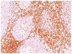
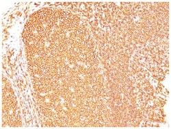
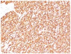
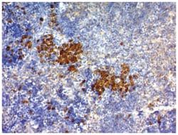
CD6 Antibody (3F7B5), Novus Biologicals™
Mouse Monoclonal Antibody
Manufacturer: Fischer Scientific
The price for this product is unavailable. Please request a quote
Antigen
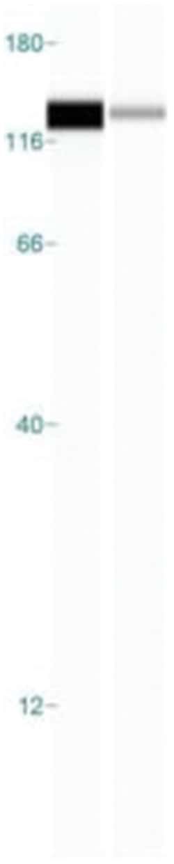
CD6
Dilution
Western Blot 0.5-1ug/ml, Flow Cytometry 0.5-1ug/million cells, Immunocytochemistry/Immunofluorescence 0.5-1ug/ml, Immunoprecipitation 0.5-1ug/500ug protein lysate, Immunohistochemistry-Paraffin 0.5-1.0ug/ml, Immunohistochemistry-Frozen 0.5-1.0ug/ml
Classification
Monoclonal
Form
Purified
Regulatory Status
RUO
Target Species
Human
Gene Accession No.
P30203
Gene ID (Entrez)
923
Immunogen
Human rheumatoid synovial T cell line ST-1
Primary or Secondary
Primary
Content And Storage
Store at 4C.
Clone
3F7B5
Applications
Western Blot, Flow Cytometry, Immunocytochemistry, Immunofluorescence, Immunoprecipitation, Immunohistochemistry (Paraffin)
Conjugate
Unconjugated
Host Species
Mouse
Research Discipline
Immunology
Formulation
PBS with 0.05% BSA. with 0.05% Sodium Azide
Gene Alias
CD6 antigenFLJ44171, CD6 molecule, T12, Tp120, TP120T-cell differentiation antigen CD6
Gene Symbols
CD6
Isotype
IgG1
Purification Method
Protein G purified
Test Specificity
CD6 is a type I transmembrane glycoprotein that contains a 24-amino acid signal sequence, three extracellular scavenger receptor cysteine-rich (SRCR) domains, a membrane-spanning domain and a 44-amino acid cytoplasmic domain. The CD6 glycoprotein is tyrosine phosphorylated during TCR-mediated T cell activation. CD6 shows significant homology to CD5. CD6 is present on mature thymocytes, peripheral T cells and a subset of B cells. Antibodies to CD6 are used to deplete T cells from bone marrow transplants to prevent graft versus host disease.
Related Products

Description
- CD6 Monoclonal specifically detects CD6 in Human samples
- It is validated for Flow Cytometry, Immunohistochemistry, Immunocytochemistry/Immunofluorescence, Immunohistochemistry-Paraffin.
Compare Similar Items
Show Difference
Antigen: CD6
Dilution: Western Blot 0.5-1ug/ml, Flow Cytometry 0.5-1ug/million cells, Immunocytochemistry/Immunofluorescence 0.5-1ug/ml, Immunoprecipitation 0.5-1ug/500ug protein lysate, Immunohistochemistry-Paraffin 0.5-1.0ug/ml, Immunohistochemistry-Frozen 0.5-1.0ug/ml
Classification: Monoclonal
Form: Purified
Regulatory Status: RUO
Target Species: Human
Gene Accession No.: P30203
Gene ID (Entrez): 923
Immunogen: Human rheumatoid synovial T cell line ST-1
Primary or Secondary: Primary
Content And Storage: Store at 4C.
Clone: 3F7B5
Applications: Western Blot, Flow Cytometry, Immunocytochemistry, Immunofluorescence, Immunoprecipitation, Immunohistochemistry (Paraffin)
Conjugate: Unconjugated
Host Species: Mouse
Research Discipline: Immunology
Formulation: PBS with 0.05% BSA. with 0.05% Sodium Azide
Gene Alias: CD6 antigenFLJ44171, CD6 molecule, T12, Tp120, TP120T-cell differentiation antigen CD6
Gene Symbols: CD6
Isotype: IgG1
Purification Method: Protein G purified
Test Specificity: CD6 is a type I transmembrane glycoprotein that contains a 24-amino acid signal sequence, three extracellular scavenger receptor cysteine-rich (SRCR) domains, a membrane-spanning domain and a 44-amino acid cytoplasmic domain. The CD6 glycoprotein is tyrosine phosphorylated during TCR-mediated T cell activation. CD6 shows significant homology to CD5. CD6 is present on mature thymocytes, peripheral T cells and a subset of B cells. Antibodies to CD6 are used to deplete T cells from bone marrow transplants to prevent graft versus host disease.
Antigen: CD63
Dilution: Western Blot 0.5-1ug/ml, Flow Cytometry 0.5-1ug/million cells, Immunocytochemistry/Immunofluorescence 0.5-1ug/ml, Immunoprecipitation 0.5-1ug/500ug protein lysate, Immunohistochemistry-Paraffin 0.5-1ug/ml, Immunohistochemistry-Frozen 0.5-1.0ug/ml
Classification: Monoclonal
Form: Purified
Regulatory Status: RUO
Target Species: Human, Mouse
Gene Accession No.: P08962
Gene ID (Entrez): 967
Immunogen: Recombinant human CD63 protein
Primary or Secondary: Primary
Content And Storage: Store at 4C.
Clone: SPM524
Applications: Western Blot, Flow Cytometry, Immunocytochemistry, Immunofluorescence, Immunoprecipitation, Immunohistochemistry (Paraffin)
Conjugate: Unconjugated
Host Species: Mouse
Research Discipline: Autophagy, Cytokine Research
Formulation: PBS with 0.05% BSA. with 0.05% Sodium Azide
Gene Alias: CD63 antigen, CD63 antigen (melanoma 1 antigen), CD63 molecule, Granulophysin, LAMP-3, Lysosomal-associated membrane protein 3, ME491, melanoma 1 antigen, Melanoma-associated antigen ME491, melanoma-associated antigen MLA1, MLA1lysosome-associated membrane glycoprotein 3, Ocular melanoma-associated antigen, OMA81H, Tetraspanin-30, tspan-30, TSPAN30granulophysin
Gene Symbols: CD63
Isotype: IgG1 κ
Purification Method: Protein A purified
Test Specificity: This product is specific for CD63
Antigen: TfR (Transferrin R)
Dilution: Western Blot 0.25-0.5ug/ml, Simple Western 10 ug/ml, Flow Cytometry 0.5-1ug/million cells, Immunocytochemistry/Immunofluorescence 1-2ug/ml, Immunohistochemistry-Paraffin 0.5-1ug/ml, Immunohistochemistry-Frozen 0.5-1.0ug/ml
Classification: Monoclonal
Form: Purified
Regulatory Status: RUO
Target Species: Human
Gene Accession No.: P02786
Gene ID (Entrez): 7037
Immunogen: KG1 acute myeloid leukemia cell line
Primary or Secondary: Primary
Content And Storage: Store at 4C.
Clone: DF1513
Applications: Western Blot, Flow Cytometry, Immunocytochemistry, Immunofluorescence, Immunohistochemistry (Paraffin)
Conjugate: Unconjugated
Host Species: Mouse
Research Discipline: Cell Biology, Cellular Markers, Hematopoietic Stem Cell Markers, Immunology, Signal Transduction, Stem Cell Markers
Formulation: PBS with 0.05% BSA. with 0.05% Sodium Azide
Gene Alias: CD71 antigen, CD71TRFR, p90, T9, TfR, TFR1, TR, transferrin receptor (p90, CD71), transferrin receptor protein 1, Trfr
Gene Symbols: TFRC
Isotype: IgG1 κ
Purification Method: Protein A purified
Test Specificity: It recognizes a ∼90-95kDa protein which is identified as cell surface transferrin receptor (CD71), a disulfide-bonded homodimeric glycoprotein of 180-190kDa (Workshop IV). This MAb is highly specific to CD71 and shows no cross-reaction with other related proteins. Ligand for transferrin receptor is the serum iron transport protein, transferrin. This receptor is broadly distributed in carcinomas, sarcomas, leukemias, and lymphomas. CD71/Transferrin receptor has been reported to be associated with cell proliferation in both normal and neoplastic tissues and useful in predicting clinical behavior or response to therapy in a number of malignancies including breast cancer.
