CD74 Antibody (BU45), Novus Biologicals™
Mouse Monoclonal Antibody
Manufacturer: Fischer Scientific
The price for this product is unavailable. Please request a quote
Antigen
CD74
Dilution
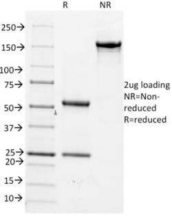
Western Blot 0.5-1ug/ml, Flow Cytometry 0.5-1ug/million cells, Immunocytochemistry/Immunofluorescence 1-2ug/ml, Immunoprecipitation 0.5-1ug/500ug protein lysate, Immunohistochemistry-Frozen 0.5-1ug/ml
Classification
Monoclonal
Form
Purified
Regulatory Status
RUO
Target Species
Human
Gene Accession No.
P044233
Gene ID (Entrez)
972
Immunogen
B lymphoblastoid cell line HFB1
Primary or Secondary
Primary
Content And Storage
Store at 4C.
Clone
BU45
Applications
Western Blot, Flow Cytometry, Immunocytochemistry, Immunofluorescence, Immunoprecipitation, Immunohistochemistry (Frozen)
Conjugate
Unconjugated
Host Species
Mouse
Research Discipline
Immunology
Formulation
PBS with 0.05% BSA. with 0.05% Sodium Azide
Gene Alias
CD74 antigen, CD74 antigen (invariant polypeptide of major histocompatibility complex, classII antigen-associated), CD74 molecule, major histocompatibility complex, class II invariant chain, DHLAGgamma chain of class II antigens, HLA class II histocompatibility antigen gamma chain, HLADG, HLA-DR antigens-associated invariant chain, HLA-DR-gamma, Ia antigen-associated invariant chain, Ia-associated invariant chain, Ia-GAMMA, Ii, MHC HLA-DR gamma chain, p33
Gene Symbols
CD74
Isotype
IgG1 κ
Purification Method
Protein A purified
Test Specificity
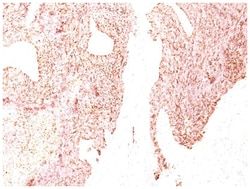
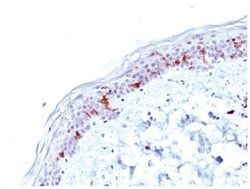
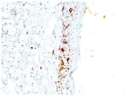
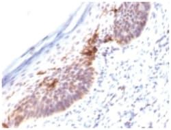
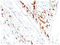
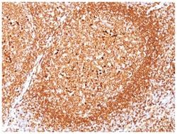
Recognizes proteins of 33, 35 and 41kDa, which are identified as various isoforms of CD74. Its epitope is localized in the extracellular domain of CD74. CD74 is a type II transmembrane protein which binds to the peptide binding groove of newly synthesized MHC class II alpha/beta heterodimers and prevents their premature association with endogenous polypeptides. The CD74 molecule plays a critical role in the presentation of peptides, by the MHC class II antigens, to CD4 positive lymphocytes. CD74 is expressed on MHC class II positive cells including B cells, a subset of activated T cells, monocytes, and dendritic cells and by various types of carcinomas. CD74 is expressed primarily by antigen presenting cells, such as B-lymphocytes (from before the pre-B cell stage to before the plasma cell stage), macrophages, and monocytes, and many epithelial cells. Anti-CD74 stains predominantly germinal center lymphocytes and B-cell lymphomas, but rarely T-cell lymphomas. Anti-CD74 has been shown to be useful in differentiating atypical fibroxanthoma (-) from malignant fibrous histiocytoma (+).
Related Products

Description
- CD74 Monoclonal specifically detects CD74 in Human samples
- It is validated for Western Blot, Flow Cytometry, Immunocytochemistry/Immunofluorescence.
Compare Similar Items
Show Difference

Antigen: CD74
Dilution: Western Blot 0.5-1ug/ml, Flow Cytometry 0.5-1ug/million cells, Immunocytochemistry/Immunofluorescence 1-2ug/ml, Immunoprecipitation 0.5-1ug/500ug protein lysate, Immunohistochemistry-Frozen 0.5-1ug/ml
Classification: Monoclonal
Form: Purified
Regulatory Status: RUO
Target Species: Human
Gene Accession No.: P044233
Gene ID (Entrez): 972
Immunogen: B lymphoblastoid cell line HFB1
Primary or Secondary: Primary
Content And Storage: Store at 4C.
Clone: BU45
Applications: Western Blot, Flow Cytometry, Immunocytochemistry, Immunofluorescence, Immunoprecipitation, Immunohistochemistry (Frozen)
Conjugate: Unconjugated
Host Species: Mouse
Research Discipline: Immunology
Formulation: PBS with 0.05% BSA. with 0.05% Sodium Azide
Gene Alias: CD74 antigen, CD74 antigen (invariant polypeptide of major histocompatibility complex, classII antigen-associated), CD74 molecule, major histocompatibility complex, class II invariant chain, DHLAGgamma chain of class II antigens, HLA class II histocompatibility antigen gamma chain, HLADG, HLA-DR antigens-associated invariant chain, HLA-DR-gamma, Ia antigen-associated invariant chain, Ia-associated invariant chain, Ia-GAMMA, Ii, MHC HLA-DR gamma chain, p33
Gene Symbols: CD74
Isotype: IgG1 κ
Purification Method: Protein A purified
Test Specificity: Recognizes proteins of 33, 35 and 41kDa, which are identified as various isoforms of CD74. Its epitope is localized in the extracellular domain of CD74. CD74 is a type II transmembrane protein which binds to the peptide binding groove of newly synthesized MHC class II alpha/beta heterodimers and prevents their premature association with endogenous polypeptides. The CD74 molecule plays a critical role in the presentation of peptides, by the MHC class II antigens, to CD4 positive lymphocytes. CD74 is expressed on MHC class II positive cells including B cells, a subset of activated T cells, monocytes, and dendritic cells and by various types of carcinomas. CD74 is expressed primarily by antigen presenting cells, such as B-lymphocytes (from before the pre-B cell stage to before the plasma cell stage), macrophages, and monocytes, and many epithelial cells. Anti-CD74 stains predominantly germinal center lymphocytes and B-cell lymphomas, but rarely T-cell lymphomas. Anti-CD74 has been shown to be useful in differentiating atypical fibroxanthoma (-) from malignant fibrous histiocytoma (+).
Antigen: CD79A
Dilution: Western Blot 0.5-1ug/ml, Flow Cytometry 0.5-1ug/million cells, Immunocytochemistry/Immunofluorescence 0.5-1ug/ml, Immunoprecipitation 0.5-1ug/500ug protein lysate, Immunohistochemistry-Paraffin 0.5-1.0ug/ml, Immunohistochemistry-Frozen 0.5-1.0ug/ml, SDS-Page
Classification: Monoclonal
Form: Purified
Regulatory Status: RUO
Target Species: Human
Gene Accession No.: P11912
Gene ID (Entrez): 973
Immunogen: A synthetic peptide corresponding to aa 202-216 (GTYQDVGSLNIADVQ) of human CD79a protein.
Primary or Secondary: Primary
Content And Storage: Store at 4C.
Clone: JCB117
Applications: Western Blot, Flow Cytometry, Immunocytochemistry, Immunofluorescence, Immunoprecipitation, Immunohistochemistry (Paraffin)
Conjugate: Unconjugated
Host Species: Mouse
Research Discipline: Adaptive Immunity, Cell Biology, Immunology, Phospho Specific
Formulation: PBS with 0.05% BSA. with 0.05% Sodium Azide
Gene Alias: CD79a antigen, CD79A antigen (immunoglobulin-associated alpha), CD79a molecule, immunoglobulin-associated alpha, IGAB-cell antigen receptor complex-associated protein alpha chain, Ig-alpha, MB1, MB-1, MB-1 membrane glycoprotein, Membrane-bound immunoglobulin-associated protein, Surface IgM-associated protein
Gene Symbols: CD79A
Isotype: IgG1 κ
Purification Method: Protein A purified
Test Specificity: A disulphide-linked heterodimer, consisting of mb-1 (or CD79a) and B29 (or CD79b) polypeptides, is non-covalently associated with membrane-bound immunoglobulins on B cells. This complex of mb-1 and B29 polypeptides and immunoglobulin constitute the B cell Ag receptor. CD79a first appears at pre B cell stage, early in maturation, and persists until the plasma cell stage where it is found as an intracellular component. CD79a is found in the majority of acute leukemias of precursor B cell type, in B cell lines, B cell lymphomas, and in some myelomas. It is not present in myeloid or T cell lines. Anti-CD79a is generally used to complement anti-CD20 especially for mature B-cell lymphomas after treatment with Rituximab (anti-CD20). This antibody will stain many of the same lymphomas as anti-CD20, but also is more likely to stain B-lymphoblastic lymphoma/leukemia than is anti-CD20. Anti-CD79a also stains more cases of plasma cell myeloma and occasionally some types of endothelial cells as wel
Antigen: Cdc20
Dilution: Western Blot 0.5-1ug/ml, Flow Cytometry 0.5-1ug/million cells, Immunocytochemistry/Immunofluorescence 0.5-1ug/ml, Immunoprecipitation 0.5-1ug/500ug protein lysate, Immunohistochemistry-Paraffin 0.5-1ug/ml, Immunohistochemistry-Frozen 0.5-1.0ug/ml, SDS-Page
Classification: Monoclonal
Form: Purified
Regulatory Status: RUO
Target Species: Human
Gene Accession No.: Q12834
Gene ID (Entrez): 991
Immunogen: Urea-denatured His6 Cdc20 human recombinant protein
Primary or Secondary: Primary
Content And Storage: Store at 4C.
Clone: AR12
Applications: Western Blot, Flow Cytometry, Immunocytochemistry, Immunofluorescence, Immunoprecipitation, Immunohistochemistry (Paraffin)
Conjugate: Unconjugated
Host Species: Mouse
Research Discipline: Cell Cycle and Replication, Core ESC Like Genes, Stem Cell Markers
Formulation: PBS with 0.05% BSA. with 0.05% Sodium Azide
Gene Alias: CDC20 cell division cycle 20 homolog, CDC20 cell division cycle 20 homolog (S. cerevisiae), cell division cycle 20 homolog (S. cerevisiae), cell division cycle protein 20 homolog, MGC102824, p55CDCS. cerevisiae, homolog)
Gene Symbols: CDC20
Isotype: IgG1 κ
Purification Method: Protein A purified
Test Specificity: Cyclins, regulatory subunits, which associate with kinases, control many of the important steps in cell cycle progression. The Cdc2 protein kinase (p34Cdc2) exhibits protein kinase activity in vitro and exists in a complex with both cyclin B and a protein homologous to p13SUC1. Cdc2 kinase is the active subunit of the M phase promoting factor (MPF) and the M phase-specific Histone H1 kinase. The p34Cdc2/cyclin B complex is required for the G2 to M transition. An additional cell cycle-dependent protein kinase, termed p55cdc, exhibits a high degree of homology with the S. cerevisiae proteins Cdc20 and Cdc4. The p55cdc transcript is readily detectable in a variety of cultured cell lines in growth phase, but disappears when cell growth is chemically arrested.