CD79A Antibody (SPM549), Novus Biologicals™
Mouse Monoclonal Antibody
Manufacturer: Fischer Scientific
The price for this product is unavailable. Please request a quote
Antigen
CD79A
Dilution
Western Blot 0.5-1ug/ml, Flow Cytometry 0.5-1ug/million cells, Immunocytochemistry/Immunofluorescence 0.5-1ug/ml, Immunoprecipitation 0.5-1ug/500ug protein lysate, Immunohistochemistry-Paraffin 0.5-1.0ug/ml, Immunohistochemistry-Frozen 0.5-1.0ug/ml
Classification
Monoclonal
Form
Purified
Regulatory Status
RUO
Target Species
Human
Gene Accession No.
P11912
Gene ID (Entrez)
973
Immunogen
A synthetic peptide corresponding to aa 202-216 (GTYQDVGSLNIADVQ) of human CD79a protein.
Primary or Secondary
Primary
Content And Storage
Store at 4C.
Molecular Weight of Antigen
44 kDa
Clone
SPM549
Applications
Western Blot, Flow Cytometry, Immunocytochemistry, Immunofluorescence, Immunoprecipitation, Immunohistochemistry (Paraffin)
Conjugate
Unconjugated
Host Species
Mouse
Research Discipline
Adaptive Immunity, Cell Biology, Immunology, Phospho Specific
Formulation
PBS with 0.05% BSA. with 0.05% Sodium Azide
Gene Alias
CD79a antigen, CD79A antigen (immunoglobulin-associated alpha), CD79a molecule, immunoglobulin-associated alpha, IGAB-cell antigen receptor complex-associated protein alpha chain, Ig-alpha, MB1, MB-1, MB-1 membrane glycoprotein, Membrane-bound immunoglobulin-associated protein, Surface IgM-associated protein
Gene Symbols
CD79A
Isotype
IgG1 κ
Purification Method
Protein A purified
Test Specificity
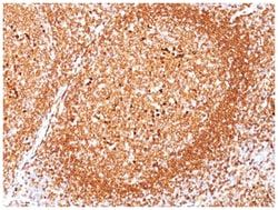
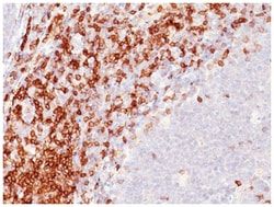
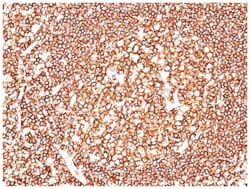
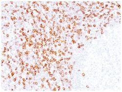
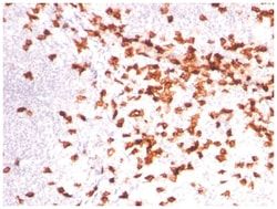
A disulphide-linked heterodimer, consisting of mb-1 (or CD79a) and B29 (or CD79b) polypeptides, is non-covalently associated with membrane-bound immunoglobulins on B cells. This complex of mb-1 and B29 polypeptides and immunoglobulin constitute the B cell Ag receptor. CD79a first appears at pre B cell stage, early in maturation, and persists until the plasma cell stage where it is found as an intracellular component. CD79a is found in the majority of acute leukemias of precursor B cell type, in B cell lines, B cell lymphomas, and in some myelomas. It is not present in myeloid or T cell lines. Anti-CD79a is generally used to complement anti-CD20 especially for mature B-cell lymphomas after treatment with Rituximab (anti-CD20). This antibody will stain many of the same lymphomas as anti-CD20, but also is more likely to stain B-lymphoblastic lymphoma/leukemia than is anti-CD20. Anti-CD79a also stains more cases of plasma cell myeloma and occasionally some types of endothelial cells as wel
Related Products


Description
- CD79A Monoclonal specifically detects CD79A in Human samples
- It is validated for Western Blot, Immunohistochemistry, Immunocytochemistry/Immunofluorescence, Immunohistochemistry-Paraffin.
Compare Similar Items
Show Difference
Antigen: CD79A
Dilution: Western Blot 0.5-1ug/ml, Flow Cytometry 0.5-1ug/million cells, Immunocytochemistry/Immunofluorescence 0.5-1ug/ml, Immunoprecipitation 0.5-1ug/500ug protein lysate, Immunohistochemistry-Paraffin 0.5-1.0ug/ml, Immunohistochemistry-Frozen 0.5-1.0ug/ml
Classification: Monoclonal
Form: Purified
Regulatory Status: RUO
Target Species: Human
Gene Accession No.: P11912
Gene ID (Entrez): 973
Immunogen: A synthetic peptide corresponding to aa 202-216 (GTYQDVGSLNIADVQ) of human CD79a protein.
Primary or Secondary: Primary
Content And Storage: Store at 4C.
Molecular Weight of Antigen: 44 kDa
Clone: SPM549
Applications: Western Blot, Flow Cytometry, Immunocytochemistry, Immunofluorescence, Immunoprecipitation, Immunohistochemistry (Paraffin)
Conjugate: Unconjugated
Host Species: Mouse
Research Discipline: Adaptive Immunity, Cell Biology, Immunology, Phospho Specific
Formulation: PBS with 0.05% BSA. with 0.05% Sodium Azide
Gene Alias: CD79a antigen, CD79A antigen (immunoglobulin-associated alpha), CD79a molecule, immunoglobulin-associated alpha, IGAB-cell antigen receptor complex-associated protein alpha chain, Ig-alpha, MB1, MB-1, MB-1 membrane glycoprotein, Membrane-bound immunoglobulin-associated protein, Surface IgM-associated protein
Gene Symbols: CD79A
Isotype: IgG1 κ
Purification Method: Protein A purified
Test Specificity: A disulphide-linked heterodimer, consisting of mb-1 (or CD79a) and B29 (or CD79b) polypeptides, is non-covalently associated with membrane-bound immunoglobulins on B cells. This complex of mb-1 and B29 polypeptides and immunoglobulin constitute the B cell Ag receptor. CD79a first appears at pre B cell stage, early in maturation, and persists until the plasma cell stage where it is found as an intracellular component. CD79a is found in the majority of acute leukemias of precursor B cell type, in B cell lines, B cell lymphomas, and in some myelomas. It is not present in myeloid or T cell lines. Anti-CD79a is generally used to complement anti-CD20 especially for mature B-cell lymphomas after treatment with Rituximab (anti-CD20). This antibody will stain many of the same lymphomas as anti-CD20, but also is more likely to stain B-lymphoblastic lymphoma/leukemia than is anti-CD20. Anti-CD79a also stains more cases of plasma cell myeloma and occasionally some types of endothelial cells as wel

Antigen: CD79A
Dilution: Western Blot 0.5-1ug/ml, Flow Cytometry 0.5-1ug/million cells, Immunocytochemistry/Immunofluorescence 0.5-1ug/ml, Immunoprecipitation 0.5-1ug/500ug protein lysate, Immunohistochemistry-Paraffin 0.5-1.0ug/ml, Immunohistochemistry-Frozen 0.5-1.0ug/ml
Classification: Monoclonal
Form: Purified
Regulatory Status: RUO
Target Species: Human, Mouse, Rat, Porcine, Bovine, Primate
Gene Accession No.: P11912
Gene ID (Entrez): 973
Immunogen: A synthetic peptide corresponding to aa 202-216 (GTYQDVGSLNIADVQ) of human CD79a protein.
Primary or Secondary: Primary
Content And Storage: Store at 4C.
Molecular Weight of Antigen: 44 kDa
Clone: SPM550
Applications: Western Blot, Flow Cytometry, Immunocytochemistry, Immunofluorescence, Immunoprecipitation, Immunohistochemistry (Paraffin)
Conjugate: Unconjugated
Host Species: Mouse
Research Discipline: Adaptive Immunity, Cell Biology, Immunology, Phospho Specific
Formulation: PBS with 0.05% BSA. with 0.05% Sodium Azide
Gene Alias: CD79a antigen, CD79A antigen (immunoglobulin-associated alpha), CD79a molecule, immunoglobulin-associated alpha, IGAB-cell antigen receptor complex-associated protein alpha chain, Ig-alpha, MB1, MB-1, MB-1 membrane glycoprotein, Membrane-bound immunoglobulin-associated protein, Surface IgM-associated protein
Gene Symbols: CD79A
Isotype: IgG1 κ
Purification Method: Protein A purified
Test Specificity: A disulphide-linked heterodimer, consisting of mb-1 (or CD79a) and B29 (or CD79b) polypeptides, is non-covalently associated with membrane-bound immunoglobulins on B cells. This complex of mb-1 and B29 polypeptides and immunoglobulin constitute the B cell Ag receptor. CD79a first appears at pre B cell stage, early in maturation, and persists until the plasma cell stage where it is found as an intracellular component. CD79a is found in the majority of acute leukemias of precursor B cell type, in B cell lines, B cell lymphomas, and in some myelomas. It is not present in myeloid or T cell lines. Anti-CD79a is generally used to complement anti-CD20 especially for mature B-cell lymphomas after treatment with Rituximab (anti-CD20). This antibody will stain many of the same lymphomas as anti-CD20, but also is more likely to stain B-lymphoblastic lymphoma/leukemia than is anti-CD20. Anti-CD79a also stains more cases of plasma cell myeloma and occasionally some types of endothelial cells as wel
Antigen: CD8 alpha
Dilution: Western Blot 0.5-1ug/ml, Flow Cytometry 0.5-1ug/million cells, Immunocytochemistry/Immunofluorescence 0.5-1ug/ml, Immunoprecipitation 1-2ug/500ug protein lysate, Immunohistochemistry-Paraffin 1-2ug/ml, Immunohistochemistry-Frozen 1-2ug/ml
Classification: Monoclonal
Form: Purified
Regulatory Status: RUO
Target Species: Human
Gene Accession No.: P01732
Gene ID (Entrez): 925
Immunogen: A 13 amino acid peptide from C-terminal cytoplasmic domain of alpha chain of human CD8 molecule.
Primary or Secondary: Primary
Content And Storage: Store at 4C.
Molecular Weight of Antigen: 32 kDa
Clone: C8/144B
Applications: Western Blot, Flow Cytometry, Immunocytochemistry, Immunofluorescence, Immunoprecipitation, Immunohistochemistry (Paraffin)
Conjugate: Unconjugated
Host Species: Mouse
Research Discipline: Adaptive Immunity, Cytokine Research, Immunology, Innate Immunity, Phospho Specific, Signal Transduction, Stem Cell Markers
Formulation: PBS with 0.05% BSA. with 0.05% Sodium Azide
Gene Alias: CD8, CD8 antigen, alpha polypeptide (p32), CD8a antigen, CD8a molecule, Leu2, Leu2 T-lymphocyte antigen, MAL, OKT8 T-cell antigen, p32, RPA-T8, RPA-T8 antibody flow, RPA-T8 CD8, RPA-T8 Clone, RPA-T8 Flow, T cell co-receptor, T8 T-cell antigen, T-cell antigen Leu2, T-cell surface glycoprotein CD8 alpha chain, T-lymphocyte differentiation antigen T8/Leu-2
Gene Symbols: CD8A
Isotype: IgG1 κ
Purification Method: Protein G purified
Test Specificity: CD8 is a cell surface receptor expressed either as a heterodimer with the CD8 beta chain (CD8 alpha/beta) or as a homodimer (CD8 alpha/alpha). A majority of thymocytes and a subpopulation of mature T cells and NK cells express CD8a. CD8 binds to MHC class 1 and through its association with protein tyrosine kinase p56lck plays a role in T cell development and activation of mature T cells. For mature T-cells, CD4 and CD8 are mutually exclusive, so anti-CD8, generally used in conjunction with anti-CD4. It is a useful marker for distinguishing helper/inducer T-lymphocytes, and most peripheral T-cell lymphomas are CD4+/CD8-. Anaplastic large cell lymphoma is usually CD4+ and CD8-, and in T-lymphoblastic lymphoma/leukemia, CD4 and CD8 are often co-expressed. CD8 is also found in littoral cell angioma of the spleen.