CD5 Antibody (SPM546), Novus Biologicals™
Mouse Monoclonal Antibody
Manufacturer: Fischer Scientific
The price for this product is unavailable. Please request a quote
Antigen
CD5
Dilution
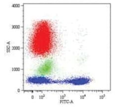
Western Blot 0.5-1ug/ml, Flow Cytometry 0.5-1ug/million cells, Immunocytochemistry/Immunofluorescence 0.5-1ug/ml, Immunoprecipitation 0.5-1ug/500ug protein lysate, Immunohistochemistry-Paraffin 0.5-1.0ug/ml, Immunohistochemistry-Frozen 0.5-1.0ug/ml
Classification
Monoclonal
Form
Purified
Regulatory Status
RUO
Target Species
Human
Gene Accession No.
P06127
Gene ID (Entrez)
921
Immunogen
Human CD5 recombinant protein
Primary or Secondary
Primary
Content And Storage
Store at 4C.
Molecular Weight of Antigen
67 kDa
Clone
SPM546
Applications
Western Blot, Flow Cytometry, Immunocytochemistry, Immunofluorescence, Immunoprecipitation, Immunohistochemistry (Paraffin)
Conjugate
Unconjugated
Host Species
Mouse
Research Discipline
Adaptive Immunity, Cell Biology, Cytokine Research, Immunology, Phospho Specific
Formulation
PBS with 0.05% BSA. with 0.05% Sodium Azide
Gene Alias
CD5 antigen, CD5 antigen (p56-62), CD5 molecule, LEU1T-cell surface glycoprotein CD5, Lymphocyte antigen T1/Leu-1, T1
Gene Symbols
CD5
Isotype
IgG1 κ
Purification Method
Protein A purified
Test Specificity
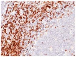
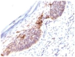
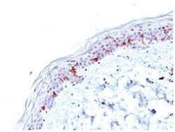
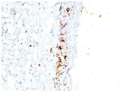
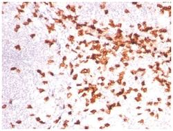
Recognizes a 67kDa transmembrane protein, which is identified as CD5. The CD5 antigen is found on 95% of thymocytes and 72% of peripheral blood lymphocytes. In lymph nodes, the main reactivity is observed in T cell areas. Anti-CD5 is a pan T-cell marker that also reacts with a range of neoplastic B-cells, e.g. chronic lymphocytic leukemia/small lymphocytic lymphoma (CLL/SLL), mantle cell lymphoma, and a subset (∼10%) of diffuse large B-cell lymphoma. CD5 aberrant expression is useful in making a diagnosis of mature T-cell neoplasms. Anti-CD5 detection is diagnostic in CLL/SLL within a panel of other B-cell markers, especially one that includes anti-CD23. Anti-CD5 is also very useful in differentiating among mature small lymphoid cell malignancies. In addition, anti-CD5 can be used in distinguishing thymic carcinoma (+) from thymoma (-). Anti-CD5 does not react with granulocytes or monocytes.
Related Products

Description
- CD5 Monoclonal specifically detects CD5 in Human samples
- It is validated for Flow Cytometry, Immunohistochemistry, Immunocytochemistry/Immunofluorescence, Immunohistochemistry-Paraffin.
Compare Similar Items
Show Difference
Antigen: CD5
Dilution: Western Blot 0.5-1ug/ml, Flow Cytometry 0.5-1ug/million cells, Immunocytochemistry/Immunofluorescence 0.5-1ug/ml, Immunoprecipitation 0.5-1ug/500ug protein lysate, Immunohistochemistry-Paraffin 0.5-1.0ug/ml, Immunohistochemistry-Frozen 0.5-1.0ug/ml
Classification: Monoclonal
Form: Purified
Regulatory Status: RUO
Target Species: Human
Gene Accession No.: P06127
Gene ID (Entrez): 921
Immunogen: Human CD5 recombinant protein
Primary or Secondary: Primary
Content And Storage: Store at 4C.
Molecular Weight of Antigen: 67 kDa
Clone: SPM546
Applications: Western Blot, Flow Cytometry, Immunocytochemistry, Immunofluorescence, Immunoprecipitation, Immunohistochemistry (Paraffin)
Conjugate: Unconjugated
Host Species: Mouse
Research Discipline: Adaptive Immunity, Cell Biology, Cytokine Research, Immunology, Phospho Specific
Formulation: PBS with 0.05% BSA. with 0.05% Sodium Azide
Gene Alias: CD5 antigen, CD5 antigen (p56-62), CD5 molecule, LEU1T-cell surface glycoprotein CD5, Lymphocyte antigen T1/Leu-1, T1
Gene Symbols: CD5
Isotype: IgG1 κ
Purification Method: Protein A purified
Test Specificity: Recognizes a 67kDa transmembrane protein, which is identified as CD5. The CD5 antigen is found on 95% of thymocytes and 72% of peripheral blood lymphocytes. In lymph nodes, the main reactivity is observed in T cell areas. Anti-CD5 is a pan T-cell marker that also reacts with a range of neoplastic B-cells, e.g. chronic lymphocytic leukemia/small lymphocytic lymphoma (CLL/SLL), mantle cell lymphoma, and a subset (∼10%) of diffuse large B-cell lymphoma. CD5 aberrant expression is useful in making a diagnosis of mature T-cell neoplasms. Anti-CD5 detection is diagnostic in CLL/SLL within a panel of other B-cell markers, especially one that includes anti-CD23. Anti-CD5 is also very useful in differentiating among mature small lymphoid cell malignancies. In addition, anti-CD5 can be used in distinguishing thymic carcinoma (+) from thymoma (-). Anti-CD5 does not react with granulocytes or monocytes.
Antigen: CD5
Dilution: Flow Cytometry 0.5-1ug/million cells, Immunocytochemistry/Immunofluorescence 0.5-1ug/ml, Immunoprecipitation 0.5-1ug/500ug protein lysate, Immunohistochemistry-Frozen 0.5-1.0ug/ml, SDS-Page
Classification: Monoclonal
Form: Purified
Regulatory Status: RUO
Target Species: Human, Primate
Gene Accession No.: P06127
Gene ID (Entrez): 921
Immunogen: Stimulated human leukocytes
Primary or Secondary: Primary
Content And Storage: Store at 4C.
Molecular Weight of Antigen: 67 kDa
Clone: CRIS-1
Applications: Flow Cytometry, Immunocytochemistry, Immunofluorescence, Immunoprecipitation, Immunohistochemistry (Frozen)
Conjugate: Unconjugated
Host Species: Mouse
Research Discipline: Adaptive Immunity, Cell Biology, Cytokine Research, Immunology, Phospho Specific
Formulation: PBS with 0.05% BSA. with 0.05% Sodium Azide
Gene Alias: CD5 antigen, CD5 antigen (p56-62), CD5 molecule, LEU1T-cell surface glycoprotein CD5, Lymphocyte antigen T1/Leu-1, T1
Gene Symbols: CD5
Isotype: IgG2a κ
Purification Method: Protein A purified
Test Specificity: Recognizes a 67kDa transmembrane protein, which is identified as CD5 (HLDA I; WS Code T 29HLDA III; WS Code T 530). The CD5 antigen is found on 95% of thymocytes and 72% of peripheral blood lymphocytes. In lymph nodes, the main reactivity is observed in T cell areas. Anti-CD5 is a pan T-cell marker that also reacts with a range of neoplastic B-cells, e.g. chronic lymphocytic leukemia/small lymphocytic lymphoma (CLL/SLL), mantle cell lymphoma, and a subset (∼10%) of diffuse large B-cell lymphoma. CD5 aberrant expression is useful in making a diagnosis of mature T-cell neoplasms.

Antigen: ICAM-3/CD50
Dilution: Western Blot 0.5-1ug/ml, Flow Cytometry 0.5-1ug/million cells, Immunocytochemistry/Immunofluorescence 0.5-1ug/ml, Immunoprecipitation 0.5-1ug/500ug protein lysate
Classification: Monoclonal
Form: Purified
Regulatory Status: RUO
Target Species: Human
Gene Accession No.: P32942
Gene ID (Entrez): 3385
Immunogen: Stimulated human leukocytes
Primary or Secondary: Primary
Content And Storage: Store at 4C.
Molecular Weight of Antigen: __
Clone: SPM505
Applications: Western Blot, Flow Cytometry, Immunocytochemistry, Immunofluorescence, Immunoprecipitation
Conjugate: Unconjugated
Host Species: Mouse
Research Discipline: Cellular Markers, Immunology
Formulation: PBS with 0.05% BSA. with 0.05% Sodium Azide
Gene Alias: CD50, CD50 antigen, CDW50CDw50, ICAM-3, ICAM-Rintercellular adhesion molecule-3, intercellular adhesion molecule 3
Gene Symbols: ICAM3
Isotype: IgG2b κ
Purification Method: Protein A purified
Test Specificity: Recognizes the D1 domain of an N-glycosylated glycoprotein of 120kDa with intra-chain disulfide bonds, identified as CD50 or ICAM-3. CD50 is the major ligand for LFA-1 (CD11a/CD18) and may have signaling role to increase adhesion. It is expressed on thymocytes and T lymphocytes and is resistant to treatment with phosphatidylinositol (PI) phospholipase C. This MAb is excellent for staining of formalin/paraffin tissues.