CD1a Antibody (O10), Novus Biologicals™
Mouse Monoclonal Antibody has been used in 1 publication
Manufacturer: Fischer Scientific
The price for this product is unavailable. Please request a quote
Antigen
CD1a
Dilution
Western Blot 0.5-1ug/ml, Flow Cytometry 0.5-1ug/million cells, Immunocytochemistry/Immunofluorescence 1-2ug/ml, Immunoprecipitation 1-2ug/500ug protein, Immunohistochemistry-Paraffin 0.5-1ug/ml, Immunohistochemistry-Frozen 0.5-1ug/ml
Classification
Monoclonal
Form
Purified
Regulatory Status
RUO
Target Species
Human
Gene Accession No.
P06126
Gene ID (Entrez)
909
Immunogen
Human thymus cells
Primary or Secondary
Primary
Content And Storage
Store at 4C.
Molecular Weight of Antigen
49 kDa
Clone
O10
Applications
Western Blot, Flow Cytometry, Immunocytochemistry, Immunofluorescence, Immunoprecipitation, Immunohistochemistry (Paraffin)
Conjugate
Unconjugated
Host Species
Mouse
Research Discipline
Dendritic Cell Markers, Immunology
Formulation
PBS with 0.05% BSA. with 0.05% Sodium Azide
Gene Alias
CD1, CD1a antigen, CD1A antigen, a polypeptide, CD1a molecule, cluster of differentiation 1 A, cortical thymocyte antigen CD1A, differentiation antigen CD1-alpha-3, epidermal dendritic cell marker CD1a, FCB6, HTA1, hTa1 thymocyte antigen, R4, T6, T-cell surface antigen T6/Leu-6, T-cell surface glycoprotein CD1a
Gene Symbols
CD1A
Isotype
IgG1 κ
Purification Method
Protein A purified
Test Specificity
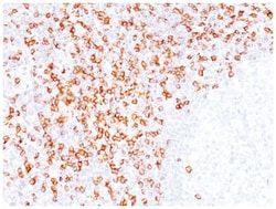
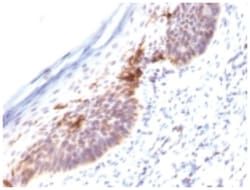
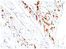
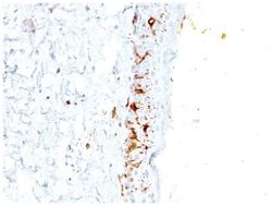
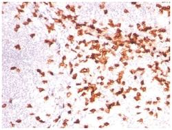
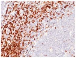
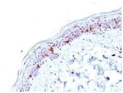
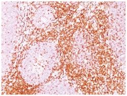
At least five CD1 genes (CD1a, b, c, d, and e) are identified. CD1 proteins have been demonstrated to restrict T cell response to non-peptide lipid and glycolipid antigens and play a role in non-classical antigen presentation. CD1a is a non-polymorphic MHC Class 1 related cell surface glycoprotein, expressed in association with Beta-2 microglobulin. Anti-CD1a labels Langerhans cell histiocytosis (Histiocytosis X), extranodal histiocytic sarcoma, a subset of T-lymphoblastic lymphoma/leukemia, and interdigitating dendritic cell sarcoma of the lymph node. When combined with antibodies against TTF-1 and CD5, anti-CD1a is useful in distinguishing between pulmonary and thymic neoplasms since CD1a is consistently expressed in thymic lymphocytes in both typical and atypical thymomas, but only focally in 1/6 of thymic carcinomas and not in lymphocytes in pulmonary neoplasms. Anti-CD1a is reported to be a new marker for perivascular epithelial cell tumor (PEComa).
Related Products
Description
- CD1a Monoclonal specifically detects CD1a in Human, Rhesus Macaque samples
- It is validated for Western Blot, Flow Cytometry, Immunohistochemistry, Immunocytochemistry/Immunofluorescence, Immunohistochemistry-Paraffin.
Compare Similar Items
Show Difference
Antigen: CD1a
Dilution: Western Blot 0.5-1ug/ml, Flow Cytometry 0.5-1ug/million cells, Immunocytochemistry/Immunofluorescence 1-2ug/ml, Immunoprecipitation 1-2ug/500ug protein, Immunohistochemistry-Paraffin 0.5-1ug/ml, Immunohistochemistry-Frozen 0.5-1ug/ml
Classification: Monoclonal
Form: Purified
Regulatory Status: RUO
Target Species: Human
Gene Accession No.: P06126
Gene ID (Entrez): 909
Immunogen: Human thymus cells
Primary or Secondary: Primary
Content And Storage: Store at 4C.
Molecular Weight of Antigen: 49 kDa
Clone: O10
Applications: Western Blot, Flow Cytometry, Immunocytochemistry, Immunofluorescence, Immunoprecipitation, Immunohistochemistry (Paraffin)
Conjugate: Unconjugated
Host Species: Mouse
Research Discipline: Dendritic Cell Markers, Immunology
Formulation: PBS with 0.05% BSA. with 0.05% Sodium Azide
Gene Alias: CD1, CD1a antigen, CD1A antigen, a polypeptide, CD1a molecule, cluster of differentiation 1 A, cortical thymocyte antigen CD1A, differentiation antigen CD1-alpha-3, epidermal dendritic cell marker CD1a, FCB6, HTA1, hTa1 thymocyte antigen, R4, T6, T-cell surface antigen T6/Leu-6, T-cell surface glycoprotein CD1a
Gene Symbols: CD1A
Isotype: IgG1 κ
Purification Method: Protein A purified
Test Specificity: At least five CD1 genes (CD1a, b, c, d, and e) are identified. CD1 proteins have been demonstrated to restrict T cell response to non-peptide lipid and glycolipid antigens and play a role in non-classical antigen presentation. CD1a is a non-polymorphic MHC Class 1 related cell surface glycoprotein, expressed in association with Beta-2 microglobulin. Anti-CD1a labels Langerhans cell histiocytosis (Histiocytosis X), extranodal histiocytic sarcoma, a subset of T-lymphoblastic lymphoma/leukemia, and interdigitating dendritic cell sarcoma of the lymph node. When combined with antibodies against TTF-1 and CD5, anti-CD1a is useful in distinguishing between pulmonary and thymic neoplasms since CD1a is consistently expressed in thymic lymphocytes in both typical and atypical thymomas, but only focally in 1/6 of thymic carcinomas and not in lymphocytes in pulmonary neoplasms. Anti-CD1a is reported to be a new marker for perivascular epithelial cell tumor (PEComa).
Antigen: CD1a
Dilution: Western Blot 0.5-1ug/ml, Flow Cytometry 0.5-1ug/million cells, Immunocytochemistry/Immunofluorescence 1-2ug/ml, Immunoprecipitation 1-2ug/500ug protein, Immunohistochemistry-Paraffin 0.5-1ug/ml, Immunohistochemistry-Frozen 0.5-1ug/ml
Classification: Monoclonal
Form: Purified
Regulatory Status: RUO
Target Species: Human
Gene Accession No.: P06126
Gene ID (Entrez): 909
Immunogen: Human thymus cells (O10); Recombinant human CD1a protein (C1A/711)
Primary or Secondary: Primary
Content And Storage: Store at 4C.
Molecular Weight of Antigen: 49 kDa
Clone: O10+C1A/711
Applications: Western Blot, Flow Cytometry, Immunocytochemistry, Immunofluorescence, Immunoprecipitation, Immunohistochemistry (Paraffin)
Conjugate: Unconjugated
Host Species: Mouse
Research Discipline: Dendritic Cell Markers, Immunology
Formulation: PBS with 0.05% BSA. with 0.05% Sodium Azide
Gene Alias: CD1, CD1a antigen, CD1A antigen, a polypeptide, CD1a molecule, cluster of differentiation 1 A, cortical thymocyte antigen CD1A, differentiation antigen CD1-alpha-3, epidermal dendritic cell marker CD1a, FCB6, HTA1, hTa1 thymocyte antigen, R4, T6, T-cell surface antigen T6/Leu-6, T-cell surface glycoprotein CD1a
Gene Symbols: CD1A
Isotype: IgG
Purification Method: Protein A purified
Test Specificity: At least five CD1 genes (CD1a, b, c, d, and e) are identified. CD1 proteins have been demonstrated to restrict T cell response to non-peptide lipid and glycolipid antigens and play a role in non-classical antigen presentation. CD1a is a non-polymorphic MHC Class 1 related cell surface glycoprotein, expressed in association with Beta-2 microglobulin. Anti-CD1a labels Langerhans cell histiocytosis (Histiocytosis X), extranodal histiocytic sarcoma, a subset of T-lymphoblastic lymphoma/leukemia, and interdigitating dendritic cell sarcoma of the lymph node. When combined with antibodies against TTF-1 and CD5, anti-CD1a is useful in distinguishing between pulmonary and thymic neoplasms since CD1a is consistently expressed in thymic lymphocytes in both typical and atypical thymomas, but only focally in 1/6 of thymic carcinomas and not in lymphocytes in pulmonary neoplasms. Anti-CD1a is reported to be a new marker for perivascular epithelial cell tumor (PEComa)
Antigen: CD6
Dilution: Western Blot 0.5-1ug/ml, Flow Cytometry 0.5-1ug/million cells, Immunocytochemistry/Immunofluorescence 0.5-1ug/ml, Immunoprecipitation 0.5-1ug/500ug protein lysate, Immunohistochemistry-Paraffin 0.5-1ug/ml, Immunohistochemistry-Frozen 0.5-1ug/ml
Classification: Monoclonal
Form: Purified
Regulatory Status: RUO
Target Species: Human
Gene Accession No.: P30203
Gene ID (Entrez): 923
Immunogen: Human recombinant CD6 protein (C6/372); Human rheumatoid synovial T cell line ST-1 (3F7B5)
Primary or Secondary: Primary
Content And Storage: Store at 4C.
Molecular Weight of Antigen: __
Clone: C6/372 + 3F7B5
Applications: Western Blot, Flow Cytometry, Immunocytochemistry, Immunofluorescence, Immunoprecipitation, Immunohistochemistry (Paraffin)
Conjugate: Unconjugated
Host Species: Mouse
Research Discipline: Immunology
Formulation: PBS with 0.05% BSA. with 0.05% Sodium Azide
Gene Alias: CD6 antigenFLJ44171, CD6 molecule, T12, Tp120, TP120T-cell differentiation antigen CD6
Gene Symbols: CD6
Isotype: IgG
Purification Method: Protein A purified
Test Specificity: CD6 is a type I transmembrane glycoprotein that contains a 24-amino acid signal sequence, three extracellular scavenger receptor cysteine-rich (SRCR) domains, a membrane-spanning domain and a 44-amino acid cytoplasmic domain. The CD6 glycoprotein is tyrosine phosphorylated during TCR-mediated T cell activation. CD6 shows significant homology to CD5. CD6 is present on mature thymocytes, peripheral T cells and a subset of B cells. Antibodies to CD6 are used to deplete T cells from bone marrow transplants to prevent graft versus host disease.