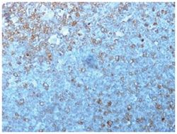
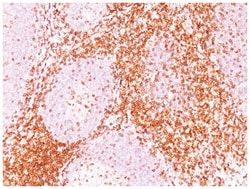
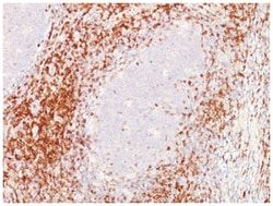
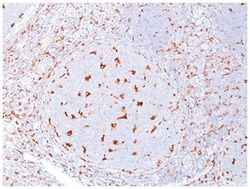
CD6 Antibody (C6/372+3F7B5), Novus Biologicals™
Mouse Monoclonal Antibody
Manufacturer: Fischer Scientific
The price for this product is unavailable. Please request a quote
Antigen
CD6
Dilution
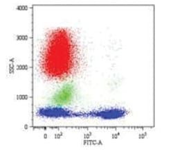
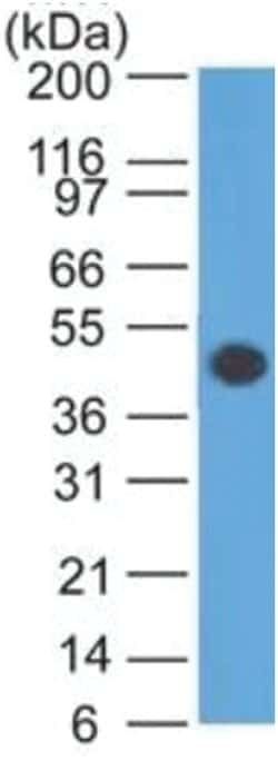
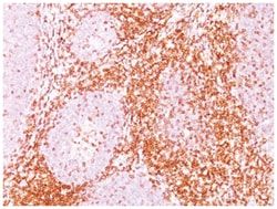
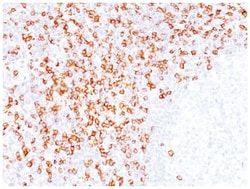
Western Blot 0.5-1ug/ml, Flow Cytometry 0.5-1ug/million cells, Immunocytochemistry/Immunofluorescence 0.5-1ug/ml, Immunoprecipitation 0.5-1ug/500ug protein lysate, Immunohistochemistry-Paraffin 0.5-1ug/ml, Immunohistochemistry-Frozen 0.5-1ug/ml
Classification
Monoclonal
Form
Purified
Regulatory Status
RUO
Target Species
Human
Gene Accession No.
P30203
Gene ID (Entrez)
923
Immunogen
Human recombinant CD6 protein (C6/372); Human rheumatoid synovial T cell line ST-1 (3F7B5)
Primary or Secondary
Primary
Content And Storage
Store at 4C.
Clone
C6/372 + 3F7B5
Applications
Western Blot, Flow Cytometry, Immunocytochemistry, Immunofluorescence, Immunoprecipitation, Immunohistochemistry (Paraffin)
Conjugate
Unconjugated
Host Species
Mouse
Research Discipline
Immunology
Formulation
PBS with 0.05% BSA. with 0.05% Sodium Azide
Gene Alias
CD6 antigenFLJ44171, CD6 molecule, T12, Tp120, TP120T-cell differentiation antigen CD6
Gene Symbols
CD6
Isotype
IgG
Purification Method
Protein A purified
Test Specificity
CD6 is a type I transmembrane glycoprotein that contains a 24-amino acid signal sequence, three extracellular scavenger receptor cysteine-rich (SRCR) domains, a membrane-spanning domain and a 44-amino acid cytoplasmic domain. The CD6 glycoprotein is tyrosine phosphorylated during TCR-mediated T cell activation. CD6 shows significant homology to CD5. CD6 is present on mature thymocytes, peripheral T cells and a subset of B cells. Antibodies to CD6 are used to deplete T cells from bone marrow transplants to prevent graft versus host disease.
Related Products


Description
- CD6 Monoclonal specifically detects CD6 in Human samples
- It is validated for Immunohistochemistry, Immunocytochemistry/Immunofluorescence, Immunohistochemistry-Paraffin.
Compare Similar Items
Show Difference
Antigen: CD6
Dilution: Western Blot 0.5-1ug/ml, Flow Cytometry 0.5-1ug/million cells, Immunocytochemistry/Immunofluorescence 0.5-1ug/ml, Immunoprecipitation 0.5-1ug/500ug protein lysate, Immunohistochemistry-Paraffin 0.5-1ug/ml, Immunohistochemistry-Frozen 0.5-1ug/ml
Classification: Monoclonal
Form: Purified
Regulatory Status: RUO
Target Species: Human
Gene Accession No.: P30203
Gene ID (Entrez): 923
Immunogen: Human recombinant CD6 protein (C6/372); Human rheumatoid synovial T cell line ST-1 (3F7B5)
Primary or Secondary: Primary
Content And Storage: Store at 4C.
Clone: C6/372 + 3F7B5
Applications: Western Blot, Flow Cytometry, Immunocytochemistry, Immunofluorescence, Immunoprecipitation, Immunohistochemistry (Paraffin)
Conjugate: Unconjugated
Host Species: Mouse
Research Discipline: Immunology
Formulation: PBS with 0.05% BSA. with 0.05% Sodium Azide
Gene Alias: CD6 antigenFLJ44171, CD6 molecule, T12, Tp120, TP120T-cell differentiation antigen CD6
Gene Symbols: CD6
Isotype: IgG
Purification Method: Protein A purified
Test Specificity: CD6 is a type I transmembrane glycoprotein that contains a 24-amino acid signal sequence, three extracellular scavenger receptor cysteine-rich (SRCR) domains, a membrane-spanning domain and a 44-amino acid cytoplasmic domain. The CD6 glycoprotein is tyrosine phosphorylated during TCR-mediated T cell activation. CD6 shows significant homology to CD5. CD6 is present on mature thymocytes, peripheral T cells and a subset of B cells. Antibodies to CD6 are used to deplete T cells from bone marrow transplants to prevent graft versus host disease.
Antigen: CD8 alpha
Dilution: Western Blot 0.5-1ug/ml, Flow Cytometry 0.5-1ug/million cells, Immunocytochemistry/Immunofluorescence 0.5-1ug/ml, Immunoprecipitation 0.5-1ug/500ug protein lysate, Immunohistochemistry-Paraffin 0.5-1ug/ml, Immunohistochemistry-Frozen 0.5-1ug/mlimmunohistochemistry-Paraffin 0.5-1ug/ml, SDS-Page
Classification: Monoclonal
Form: Purified
Regulatory Status: RUO
Target Species: Human
Gene Accession No.: P01732
Gene ID (Entrez): 925
Immunogen: Human CD8 recombinant protein (C8/468); A 13 amino acid peptide from C-terminal cytoplasmic domain of alpha chain of human CD8 molecule (C8/144B)
Primary or Secondary: Primary
Content And Storage: Store at 4C.
Clone: C8/468 + C8/144B
Applications: Western Blot, Flow Cytometry, Immunocytochemistry, Immunofluorescence, Immunoprecipitation, Immunohistochemistry (Paraffin)
Conjugate: Unconjugated
Host Species: Mouse
Research Discipline: Adaptive Immunity, Cytokine Research, Immunology, Innate Immunity, Signal Transduction, Stem Cell Markers
Formulation: PBS with 0.05% BSA. with 0.05% Sodium Azide
Gene Alias: CD8, CD8 antigen, alpha polypeptide (p32), CD8a antigen, CD8a molecule, Leu2, Leu2 T-lymphocyte antigen, MAL, OKT8 T-cell antigen, p32, RPA-T8, RPA-T8 antibody flow, RPA-T8 CD8, RPA-T8 Clone, RPA-T8 Flow, T cell co-receptor, T8 T-cell antigen, T-cell antigen Leu2, T-cell surface glycoprotein CD8 alpha chain, T-lymphocyte differentiation antigen T8/Leu-2
Gene Symbols: CD8A
Isotype: IgG
Purification Method: Protein A purified
Test Specificity: CD8 is a cell surface receptor expressed either as a heterodimer with the CD8 beta chain (CD8 alpha/beta) or as a homodimer (CD8 alpha/alpha). A majority of thymocytes and a subpopulation of mature T cells and NK cells express CD8a. CD8 binds to MHC class 1 and through its association with protein tyrosine kinase p56lck plays a role in T cell development and activation of mature T cells. For mature T-cells, CD4 and CD8 are mutually exclusive, so anti-CD8, generally used in conjunction with anti-CD4. It is a useful marker for distinguishing helper/inducer T-lymphocytes, and most peripheral T-cell lymphomas are CD4+/CD8-. Anaplastic large cell lymphoma is usually CD4+ and CD8-, and in T-lymphoblastic lymphoma/leukemia, CD4 and CD8 are often co-expressed. CD8 is also found in littoral cell angioma of the spleen.