GFAP Mouse, Clone: SPM507, Novus Biologicals™
Mouse Monoclonal Antibody
Manufacturer: Fischer Scientific
The price for this product is unavailable. Please request a quote
Antigen
GFAP
Dilution
Western Blot 0.5-1ug/ml, Flow Cytometry 0.5-1ug/million cells, Immunocytochemistry/Immunofluorescence 1-2ug/ml, Immunoprecipitation 1-2ug/500ug, Immunohistochemistry-Paraffin 0.5-1ug/ml, Immunohistochemistry-Frozen 0.5-1.0ug/ml
Classification
Monoclonal
Form
Purified
Regulatory Status
RUO
Target Species
Human, Mouse, Rat, Porcine, Bovine, Chicken, Rabbit
Gene Accession No.
P14136
Gene ID (Entrez)
2670
Immunogen
GFAP isolated from pig spinal cord
Primary or Secondary
Primary
Content And Storage
Store at 4C.
Molecular Weight of Antigen
50 kDa
Clone
SPM507
Applications
Western Blot, Flow Cytometry, Immunocytochemistry, Immunofluorescence, Immunoprecipitation, Immunohistochemistry (Paraffin)
Conjugate
Unconjugated
Host Species
Mouse
Research Discipline
Astrocyte Markers, Cancer, Cellular Markers, Cytoskeleton Markers, Neuronal Stem Cell Markers, Neuroscience, Stem Cell Markers
Formulation
PBS with 0.05% BSA. with 0.05% Sodium Azide
Gene Alias
FLJ45472, GFAP astrocytes, glial fibrillary acidic protein
Gene Symbols
GFAP
Isotype
IgG1
Purification Method
Protein G purified
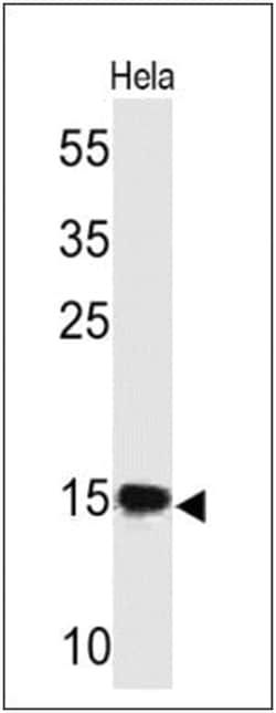
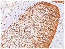
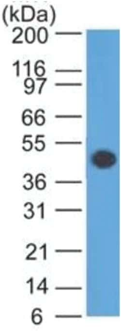
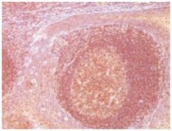
Test Specificity
This MAb recognizes a protein of ∼50kDa which is identified as Glial Fibrillary Acidic Protein (GFAP). It shows no cross-reaction with other intermediate filament proteins. GFAP is specifically found in astroglia. GFAP is a very popular marker for localizing benign astrocyte and neoplastic cells of glial origin in the central nervous system. Antibody to GFAP is useful in differentiating primary gliomas from metastatic lesions in the brain and for documenting astrocytic differentiation in tumors outside the CNS.
Related Products


Description
- GFAP Monoclonal specifically detects GFAP in Human, Mouse, Rat, Porcine, Bovine, Chicken, Rabbit samples
- It is validated for Western Blot, Flow Cytometry, Immunohistochemistry, Immunocytochemistry/Immunofluorescence, Immunohistochemistry-Paraffin, Flow (Intracellular).
Compare Similar Items
Show Difference
Antigen: GFAP
Dilution: Western Blot 0.5-1ug/ml, Flow Cytometry 0.5-1ug/million cells, Immunocytochemistry/Immunofluorescence 1-2ug/ml, Immunoprecipitation 1-2ug/500ug, Immunohistochemistry-Paraffin 0.5-1ug/ml, Immunohistochemistry-Frozen 0.5-1.0ug/ml
Classification: Monoclonal
Form: Purified
Regulatory Status: RUO
Target Species: Human, Mouse, Rat, Porcine, Bovine, Chicken, Rabbit
Gene Accession No.: P14136
Gene ID (Entrez): 2670
Immunogen: GFAP isolated from pig spinal cord
Primary or Secondary: Primary
Content And Storage: Store at 4C.
Molecular Weight of Antigen: 50 kDa
Clone: SPM507
Applications: Western Blot, Flow Cytometry, Immunocytochemistry, Immunofluorescence, Immunoprecipitation, Immunohistochemistry (Paraffin)
Conjugate: Unconjugated
Host Species: Mouse
Research Discipline: Astrocyte Markers, Cancer, Cellular Markers, Cytoskeleton Markers, Neuronal Stem Cell Markers, Neuroscience, Stem Cell Markers
Formulation: PBS with 0.05% BSA. with 0.05% Sodium Azide
Gene Alias: FLJ45472, GFAP astrocytes, glial fibrillary acidic protein
Gene Symbols: GFAP
Isotype: IgG1
Purification Method: Protein G purified
Test Specificity: This MAb recognizes a protein of ∼50kDa which is identified as Glial Fibrillary Acidic Protein (GFAP). It shows no cross-reaction with other intermediate filament proteins. GFAP is specifically found in astroglia. GFAP is a very popular marker for localizing benign astrocyte and neoplastic cells of glial origin in the central nervous system. Antibody to GFAP is useful in differentiating primary gliomas from metastatic lesions in the brain and for documenting astrocytic differentiation in tumors outside the CNS.
Antigen: Bcl-10
Dilution: Western Blot 0.5-1ug/ml, Flow Cytometry 0.5-1ug/million cells, Immunocytochemistry/Immunofluorescence 1-2ug/ml, Immunoprecipitation 1-2ug/500ug, Immunohistochemistry-Paraffin 0.5-1ug/ml, Immunohistochemistry-Frozen 0.5-1.0ug/ml
Classification: Monoclonal
Form: Purified
Regulatory Status: RUO
Target Species: Human
Gene Accession No.: O95999
Gene ID (Entrez): 8915
Immunogen: Human BCL10 recombinant protein (epitope aa122-168)
Primary or Secondary: Primary
Content And Storage: Store at 4C.
Molecular Weight of Antigen: 33 kDa
Clone: SPM520
Applications: Western Blot, Flow Cytometry, Immunocytochemistry, Immunofluorescence, Immunoprecipitation, Immunohistochemistry (Paraffin)
Conjugate: Unconjugated
Host Species: Mouse
Research Discipline: Apoptosis, Cancer
Formulation: PBS with 0.05% BSA. with 0.05% Sodium Azide
Gene Alias: B-cell CLL/lymphoma 10CIPERCLAPB-cell lymphoma/leukemia 10, Bcl-10, CARD-containing apoptotic signaling protein, CARD-containing molecule enhancing NF-kappa-B, CARD-containing proapoptotic protein, CARD-like apoptotic protein, CARMEN, caspase-recruiting domain-containing protein, cCARMEN, c-E10bcl-10, CED-3/ICH-1 prodomain homologous E10-like regulator, Cellular homolog of vCARMEN, cellular-E10, hCLAP, Mammalian CARD-containing adapter molecule E10, mE10CARD containing molecule enhancing NF-kB
Gene Symbols: BCL10
Isotype: IgG1 κ
Purification Method: Protein A purified
Test Specificity: This MAb labels subpopulations of normal B and T cells and is a useful tool for the sub-classification of lymphomas.
Antigen: Cytokeratin 19
Dilution: Western Blot 0.5-1ug/ml, Flow Cytometry 0.5-1ug/million cells, Immunocytochemistry/Immunofluorescence 1-2ug/ml, Immunoprecipitation 1-2ug/500ug, Immunohistochemistry-Paraffin 0.5-1ug/ml, Immunohistochemistry-Frozen 0.5-1.0ug/ml
Classification: Monoclonal
Form: Purified
Regulatory Status: RUO
Target Species: Human, Mouse
Gene Accession No.: P08727
Gene ID (Entrez): 3880
Immunogen: Human mammary epithelial organoids
Primary or Secondary: Primary
Content And Storage: Store at 4C.
Molecular Weight of Antigen: 40 kDa
Clone: SPM561
Applications: Western Blot, Flow Cytometry, Immunocytochemistry, Immunofluorescence, Immunoprecipitation, Immunohistochemistry (Paraffin)
Conjugate: Unconjugated
Host Species: Mouse
Research Discipline: Cancer, Cell Biology, Cellular Markers, Cytoskeleton Markers, Neuroscience, Stem Cell Markers
Formulation: PBS with 0.05% BSA. with 0.05% Sodium Azide
Gene Alias: CK19, CK-19, cytokeratin 19, Cytokeratin-19,40-kDa keratin intermediate filament, K19cytokeratin-19, K1CS, keratin 19, keratin, type I cytoskeletal 19, keratin, type I, 40-kd, keratin-19, MGC15366
Gene Symbols: KRT19
Isotype: IgG1 κ
Purification Method: Protein A purified
Test Specificity: This Ab reacts with the rod domain of human cytokeratin 19 (CK19), a polypeptide of 40kDa. CK19 is expressed in sweat gland, mammary gland ductal and secretory cells, bile ducts, gastrointestinal tract, bladder urothelium, oral epithelia, esophagus, and ectocervical epithelium. Anti-CK19 reacts with a wide variety of epithelial malignancies including adenocarcinomas of the colon, stomach, pancreas, biliary tract, liver, and breast. Perhaps the most useful application is the identification of thyroid carcinoma of the papillary type, although 50%-60% of follicular carcinomas are also labeled. Anti-CK19 is a useful marker for detection of tumor cells in lymph nodes, peripheral blood, bone marrow and breast cancer.