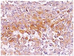
Fibronectin/Anastellin Mouse, Clone: SPM246, Novus Biologicals™
Mouse Monoclonal Antibody
Manufacturer: Fischer Scientific
The price for this product is unavailable. Please request a quote
Antigen
Fibronectin
Dilution
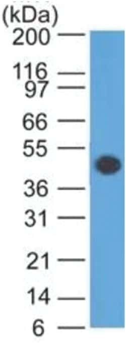
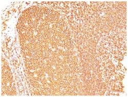
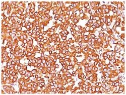
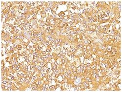
Western Blot 0.5-1ug/ml, Flow Cytometry 0.5-1ug/million cells, Immunocytochemistry/Immunofluorescence 0.5-1ug/ml, Immunoprecipitation 0.5-1ug/500ug protein lysate, Immunohistochemistry-Paraffin 0.5-1.0ug/ml, Immunohistochemistry-Frozen 0.5-1.0ug/ml
Classification
Monoclonal
Form
Purified
Regulatory Status
RUO
Target Species
Human, Mouse, Rat, Porcine
Gene Accession No.
P02751
Gene ID (Entrez)
2335
Immunogen
T-cell lymphoma biopsy
Primary or Secondary
Primary
Content And Storage
Store at 4C.
Clone
SPM246
Applications
Western Blot, Flow Cytometry, Immunocytochemistry, Immunofluorescence, Immunoprecipitation, Immunohistochemistry (Paraffin)
Conjugate
Unconjugated
Host Species
Mouse
Research Discipline
Cancer, Cellular Markers, Extracellular Matrix, Mesenchymal Stem Cell Markers, Neuronal Stem Cell Markers, Stem Cell Markers
Formulation
PBS with 0.05% BSA. with 0.05% Sodium Azide
Gene Alias
CIGDKFZp686O13149, Cold-insoluble globulin, DKFZp686F10164, DKFZp686H0342, DKFZp686I1370, fibronectin, fibronectin 1, FNFINC, FNZ, GFND2ED-B, LETSGFND, migration-stimulating factor, MSF
Gene Symbols
FN1
Isotype
IgG1 κ
Purification Method
Protein A purified
Test Specificity
Fibronectin is a soluble dimeric glycoprotein of 440kDa, which is present in cells, extracellular matrix, and blood. This MAb reacts with the cellular as well as plasma form of fibronectin. Reportedly, after iv administration, this MAb localizes to tumor vessels where it binds to the underlying basement. Epitope recognized by this antibody is not accessible in normal tissues to the circulating MAb indicating that it can be used to specifically target tumor vessels in vivo. TV-1 is reportedly useful for delivering vasoactive agents to tumors to induce increased vascular permeability or blood flow prior to treatment with chemotherapeutic drugs or MAbs.
Related Products


Description
- Fibronectin Monoclonal specifically detects Fibronectin in Human, Mouse, Rat, Porcine samples
- It is validated for Flow Cytometry, Immunohistochemistry, Immunocytochemistry/Immunofluorescence, Immunohistochemistry-Paraffin.
Compare Similar Items
Show Difference
Antigen: Fibronectin
Dilution: Western Blot 0.5-1ug/ml, Flow Cytometry 0.5-1ug/million cells, Immunocytochemistry/Immunofluorescence 0.5-1ug/ml, Immunoprecipitation 0.5-1ug/500ug protein lysate, Immunohistochemistry-Paraffin 0.5-1.0ug/ml, Immunohistochemistry-Frozen 0.5-1.0ug/ml
Classification: Monoclonal
Form: Purified
Regulatory Status: RUO
Target Species: Human, Mouse, Rat, Porcine
Gene Accession No.: P02751
Gene ID (Entrez): 2335
Immunogen: T-cell lymphoma biopsy
Primary or Secondary: Primary
Content And Storage: Store at 4C.
Clone: SPM246
Applications: Western Blot, Flow Cytometry, Immunocytochemistry, Immunofluorescence, Immunoprecipitation, Immunohistochemistry (Paraffin)
Conjugate: Unconjugated
Host Species: Mouse
Research Discipline: Cancer, Cellular Markers, Extracellular Matrix, Mesenchymal Stem Cell Markers, Neuronal Stem Cell Markers, Stem Cell Markers
Formulation: PBS with 0.05% BSA. with 0.05% Sodium Azide
Gene Alias: CIGDKFZp686O13149, Cold-insoluble globulin, DKFZp686F10164, DKFZp686H0342, DKFZp686I1370, fibronectin, fibronectin 1, FNFINC, FNZ, GFND2ED-B, LETSGFND, migration-stimulating factor, MSF
Gene Symbols: FN1
Isotype: IgG1 κ
Purification Method: Protein A purified
Test Specificity: Fibronectin is a soluble dimeric glycoprotein of 440kDa, which is present in cells, extracellular matrix, and blood. This MAb reacts with the cellular as well as plasma form of fibronectin. Reportedly, after iv administration, this MAb localizes to tumor vessels where it binds to the underlying basement. Epitope recognized by this antibody is not accessible in normal tissues to the circulating MAb indicating that it can be used to specifically target tumor vessels in vivo. TV-1 is reportedly useful for delivering vasoactive agents to tumors to induce increased vascular permeability or blood flow prior to treatment with chemotherapeutic drugs or MAbs.
Antigen: PMEL17/SILV
Dilution: Western Blot 0.5-1.0ug/ml, Flow Cytometry 0.5-1ug/million cells, Immunocytochemistry/Immunofluorescence 0.5-1ug/ml, Immunoprecipitation 0.5-1ug/500ug protein lysate, Immunohistochemistry-Paraffin 0.5-1.0ug/ml, Immunohistochemistry-Frozen 0.5-1.0ug/ml
Classification: Monoclonal
Form: Purified
Regulatory Status: RUO
Target Species: Human, Canine (Negative), Rat (Negative)
Gene Accession No.: P40967
Gene ID (Entrez): 6490
Immunogen: Extract of pigmented melanoma metastases from lymph nodes
Primary or Secondary: Primary
Content And Storage: Store at 4C.
Clone: SPM142
Applications: Western Blot, Flow Cytometry, Immunocytochemistry, Immunofluorescence, Immunoprecipitation, Immunohistochemistry (Paraffin)
Conjugate: Unconjugated
Host Species: Mouse
Research Discipline: __
Formulation: PBS with 0.05% BSA. with 0.05% Sodium Azide
Gene Alias: D12S53EP1, gp100, ME20, ME20-M, melanocyte protein mel 17, Melanocyte protein Pmel 17, Melanocytes lineage-specific antigen GP100, Melanoma-associated ME20 antigen, melanosomal matrix protein17, PMEL17P100, premelanosome proteinME20M, SI, SIL, silver (mouse homolog) like, silver homolog (mouse), Silver locus protein homolog, silver, mouse, homolog of, SILVPmel17
Gene Symbols: PMEL
Isotype: IgG1 κ
Purification Method: Protein A purified
Test Specificity: By immunohistochemistry, it specifically recognizes a protein in melanocytes and melanomas. This MAb reacts with junctional and blue nevus cells and variably with fetal and neonatal melanocytes. Intradermal nevi, normal adult melanocytes, and non-melanocytic cells are negative. It does not stain tumor cells of epithelial, lymphoid, glial, or mesenchymal origin. Metastatic amelanotic melanoma can often be confused with a variety of poorly differentiated carcinomas, large cell lymphomas, and sarcomas using H & E stains alone. It is also difficult to differentiate melanoma from spindle cell carcinomas and various types of mesenchymal neoplasms. It stains fetal and neonatal melanocytes, junctional and blue nevus cells, and malignant melanoma. This MAb also stains Angiomyolipoma.
Antigen: PMEL17/SILV
Dilution: Flow Cytometry 0.5-1ug/million cells, Immunocytochemistry/Immunofluorescence 1-2ug/ml, Immunohistochemistry-Paraffin 0.5-1.0ug/ml, Immunohistochemistry-Frozen 0.5-1.0ug/ml
Classification: Monoclonal
Form: Purified
Regulatory Status: RUO
Target Species: Human, Equine, Rat (Negative)
Gene Accession No.: P40967
Gene ID (Entrez): 6490
Immunogen: Membranes from a human melanoma metastasis
Primary or Secondary: Primary
Content And Storage: Store at 4C.
Clone: SPM286
Applications: Flow Cytometry, Immunocytochemistry, Immunofluorescence, Immunohistochemistry (Paraffin), Immunohistochemistry (Frozen)
Conjugate: Unconjugated
Host Species: Mouse
Research Discipline: __
Formulation: PBS with 0.05% BSA. with 0.05% Sodium Azide
Gene Alias: D12S53EP1, gp100, ME20, ME20-M, melanocyte protein mel 17, Melanocyte protein Pmel 17, Melanocytes lineage-specific antigen GP100, Melanoma-associated ME20 antigen, melanosomal matrix protein17, PMEL17P100, premelanosome proteinME20M, SI, SIL, silver (mouse homolog) like, silver homolog (mouse), Silver locus protein homolog, silver, mouse, homolog of, SILVPmel17
Gene Symbols: PMEL
Isotype: IgG2b κ
Purification Method: Protein A purified
Test Specificity: By immunohistochemistry, it specifically recognizes a protein in melanocytes and melanomas. This MAb reacts with junctional and blue nevus cells and variably with fetal and neonatal melanocytes. Intradermal nevi, normal adult melanocytes, and non-melanocytic cells are negative. It does not stain tumor cells of epithelial, lymphoid, glial, or mesenchymal origin.This Mab labels formalin-fixed, paraffin-embedded melanomas and other tumors showing melanocytic differentiation.