PCNA Mouse, Clone: SPM350, Novus Biologicals™
Mouse Monoclonal Antibody
Manufacturer: Fischer Scientific
The price for this product is unavailable. Please request a quote
Antigen
PCNA
Dilution
Western Blot 0.5-1ug/ml, Flow Cytometry 0.5-1ug/million cells, Immunocytochemistry/Immunofluorescence 0.5-1ug/ml, Immunoprecipitation 0.5-1ug/500ug protein lysate, Immunohistochemistry-Paraffin 0.5-1.0ug/ml, Immunohistochemistry-Frozen 0.5-1.0ug/ml
Classification
Monoclonal
Form
Purified
Regulatory Status
RUO
Target Species
Human, Mouse, Rat, Porcine, Chicken, Drosophila, Primate, Yeast, Zebrafish
Gene Accession No.
P12004
Gene ID (Entrez)
5111
Immunogen
Rat PCNA/Protein A fusion protein
Primary or Secondary
Primary
Content And Storage
Store at 4C.
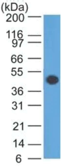
Molecular Weight of Antigen
36 kDa
Clone
SPM350
Applications
Western Blot, Flow Cytometry, Immunocytochemistry, Immunofluorescence, Immunoprecipitation, Immunohistochemistry (Paraffin)
Conjugate
Unconjugated
Host Species
Mouse
Research Discipline
Autophagy, Base Excision Repair, Cell Cycle and Replication, Cellular Markers, Core ESC Like Genes, DNA Polymerases, DNA Repair, Phospho Specific, Stem Cell Markers
Formulation
PBS with 0.05% BSA. with 0.05% Sodium Azide
Gene Alias
cyclin, DNA polymerase delta auxiliary protein, MGC8367, proliferating cell nuclear antigen
Gene Symbols
PCNA
Isotype
IgG2a κ
Purification Method
Protein A purified
Test Specificity
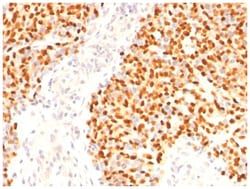
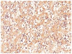
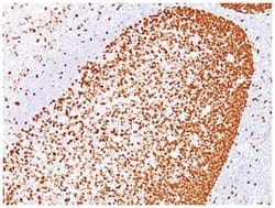
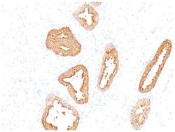
Recognizes a non-histone protein of 36kDa, which is identified as proliferating cell nuclear antigen (PCNA). It is also known as cyclin or polymerase delta auxiliary protein. Elevated expression of PCNA/cyclin has been shown in the nucleus during late G1 phase immediately before the onset of DNA synthesis, becoming maximal during S-phase and declining during G2 and M phases. This MAb is excellent for multiple applications.
Related Products

Description
- PCNA Monoclonal specifically detects PCNA in Human, Mouse, Rat, Porcine, Chicken, Drosophila, Monkey, S
- pombe, Yeast, Zebrafish samples
- It is validated for Western Blot, Flow Cytometry, Immunohistochemistry, Immunocytochemistry/Immunofluorescence, Immunohistochemistry-Paraffin.
Compare Similar Items
Show Difference
Antigen: PCNA
Dilution: Western Blot 0.5-1ug/ml, Flow Cytometry 0.5-1ug/million cells, Immunocytochemistry/Immunofluorescence 0.5-1ug/ml, Immunoprecipitation 0.5-1ug/500ug protein lysate, Immunohistochemistry-Paraffin 0.5-1.0ug/ml, Immunohistochemistry-Frozen 0.5-1.0ug/ml
Classification: Monoclonal
Form: Purified
Regulatory Status: RUO
Target Species: Human, Mouse, Rat, Porcine, Chicken, Drosophila, Primate, Yeast, Zebrafish
Gene Accession No.: P12004
Gene ID (Entrez): 5111
Immunogen: Rat PCNA/Protein A fusion protein
Primary or Secondary: Primary
Content And Storage: Store at 4C.
Molecular Weight of Antigen: 36 kDa
Clone: SPM350
Applications: Western Blot, Flow Cytometry, Immunocytochemistry, Immunofluorescence, Immunoprecipitation, Immunohistochemistry (Paraffin)
Conjugate: Unconjugated
Host Species: Mouse
Research Discipline: Autophagy, Base Excision Repair, Cell Cycle and Replication, Cellular Markers, Core ESC Like Genes, DNA Polymerases, DNA Repair, Phospho Specific, Stem Cell Markers
Formulation: PBS with 0.05% BSA. with 0.05% Sodium Azide
Gene Alias: cyclin, DNA polymerase delta auxiliary protein, MGC8367, proliferating cell nuclear antigen
Gene Symbols: PCNA
Isotype: IgG2a κ
Purification Method: Protein A purified
Test Specificity: Recognizes a non-histone protein of 36kDa, which is identified as proliferating cell nuclear antigen (PCNA). It is also known as cyclin or polymerase delta auxiliary protein. Elevated expression of PCNA/cyclin has been shown in the nucleus during late G1 phase immediately before the onset of DNA synthesis, becoming maximal during S-phase and declining during G2 and M phases. This MAb is excellent for multiple applications.
Antigen: Kallikrein 3/PSA
Dilution: Western Blot 0.5-1.0ug/ml, Flow Cytometry 0.5-1ug/million cells, Immunocytochemistry/Immunofluorescence 0.5-1ug/ml, Immunoprecipitation 0.5-1ug/500ug protein lysate, Immunohistochemistry-Paraffin 0.5-1.0ug/ml, Immunohistochemistry-Frozen 0.5-1.0ug/ml, SDS-Page
Classification: Monoclonal
Form: Purified
Regulatory Status: RUO
Target Species: Human
Gene Accession No.: P07288
Gene ID (Entrez): 354
Immunogen: PSA (p30) from human sperm plasma
Primary or Secondary: Primary
Content And Storage: Store at 4C.
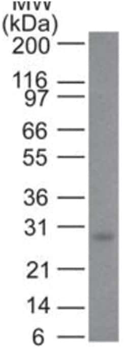
Molecular Weight of Antigen: __
Clone: A67-B/E3
Applications: Western Blot, Flow Cytometry, Immunocytochemistry, Immunofluorescence, Immunoprecipitation, Immunohistochemistry (Paraffin)
Conjugate: Unconjugated
Host Species: Mouse
Research Discipline: Cancer, Cellular Markers, Prostate Cancer
Formulation: PBS with 0.05% BSA. with 0.05% Sodium Azide
Gene Alias: APS seminin, EC 3.4.21, EC 3.4.21.77, Gamma-seminoprotein, hK3, kallikrein 3, (prostate specific antigen), Kallikrein-3, kallikrein-related peptidase 3, KLK2A1, P-30 antigen, prostate specific antigen, prostate-specific antigen, PSA, PSA semenogelase, Semenogelase, Seminin
Gene Symbols: KLK3
Isotype: IgG1 κ
Purification Method: Protein A purified
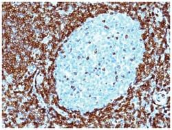
Test Specificity: Recognizes a single protein of 33-34kDa, identified as the prostate specific antigen (PSA). This MAb is highly specific to PSA and stains prostatic secretory and ductal epithelium in both normal and neoplastic tissues. PSA is a chymotrypsin-like serine protease (kallikrein family) exclusively produced by the prostate epithelium, and abundant in seminal fluid. PSA can be detected in the sera of patients with prostatic carcinoma. It is predominantly complexed to a liver-derived serine protease inhibitor, alpha-1-antichymotrypsin (ACT). A higher proportion of serum PSA is complexed to ACT in prostate cancer than in benign prostate hyperplasia. This MAb makes an excellent pair with MAb 1A7G6B6 for PSA tests.
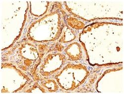
Antigen: Thyroglobulin
Dilution: Western Blot 0.5-1ug/ml, Flow Cytometry 0.5-1ug/million cells, Immunohistochemistry-Paraffin 0.5-1.0ug/ml
Classification: Monoclonal
Form: Purified
Regulatory Status: RUO
Target Species: Human, Mouse, Rat
Gene Accession No.: P01266
Gene ID (Entrez): 7038
Immunogen: Human thyroid follicular cells
Primary or Secondary: Primary
Content And Storage: Store at 4C.
Molecular Weight of Antigen: __
Clone: SPM517
Applications: Western Blot, Flow Cytometry, Immunohistochemistry (Paraffin)
Conjugate: Unconjugated
Host Species: Mouse
Research Discipline: Cancer, Cell Biology
Formulation: PBS with 0.05% BSA. with 0.05% Sodium Azide
Gene Alias: AITD3TGN, TDH3, Tg, thyroglobulin
Gene Symbols: TG
Isotype: IgG1 κ
Purification Method: Protein A purified
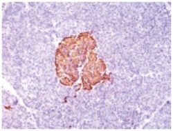
Test Specificity: Thyroglobulin is a 660kDa dimeric pre-protein with mutiple glycosylation sites. It is produced by and processed within the thyroid gland to produce the hormone thyroxine and triiodothyronine. Prior to forming dimers, thyroglobulin monomers undergo conformational maturation in the endoplasmic reticulation. The vast majority of follicular carcinomas of the thyroid will give positive immunoreactivity for anti-thyroglobulin even though sometimes only focally. Poorly differentiated carcinomas of the thyroid are frequently anti-thyroglobulin negative. Adenocarcinomas of other-than-thyroid origin do not react with this antibody. This antibody is useful in identification of thyroid carcinoma of the papillary and follicular types. Presence of thyroglobulin in metastatic lesions establishes the thyroid origin of tumor. Anti-thyroglobulin, combined with anti-calcitonin, can identify medullary carcinomas of the thyroid. Furthermore, anti-thyroglobulin, combined with anti-TTF1, can be a reliable ma
