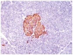
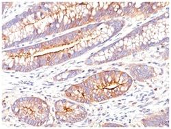
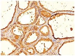
Thyroglobulin Mouse, Clone: SPM517, Novus Biologicals™
Mouse Monoclonal Antibody
Manufacturer: Fischer Scientific
The price for this product is unavailable. Please request a quote
Antigen
Thyroglobulin
Dilution
Western Blot 0.5-1ug/ml, Flow Cytometry 0.5-1ug/million cells, Immunohistochemistry-Paraffin 0.5-1.0ug/ml
Classification
Monoclonal
Form
Purified
Regulatory Status
RUO
Target Species
Human, Mouse, Rat
Gene Accession No.
P01266
Gene ID (Entrez)
7038
Immunogen
Human thyroid follicular cells
Primary or Secondary
Primary
Content And Storage
Store at 4C.
Clone
SPM517
Applications
Western Blot, Flow Cytometry, Immunohistochemistry (Paraffin)
Conjugate
Unconjugated
Host Species
Mouse
Research Discipline
Cancer, Cell Biology
Formulation
PBS with 0.05% BSA. with 0.05% Sodium Azide
Gene Alias
AITD3TGN, TDH3, Tg, thyroglobulin
Gene Symbols
TG
Isotype
IgG1 κ
Purification Method
Protein A purified
Test Specificity
Thyroglobulin is a 660kDa dimeric pre-protein with mutiple glycosylation sites. It is produced by and processed within the thyroid gland to produce the hormone thyroxine and triiodothyronine. Prior to forming dimers, thyroglobulin monomers undergo conformational maturation in the endoplasmic reticulation. The vast majority of follicular carcinomas of the thyroid will give positive immunoreactivity for anti-thyroglobulin even though sometimes only focally. Poorly differentiated carcinomas of the thyroid are frequently anti-thyroglobulin negative. Adenocarcinomas of other-than-thyroid origin do not react with this antibody. This antibody is useful in identification of thyroid carcinoma of the papillary and follicular types. Presence of thyroglobulin in metastatic lesions establishes the thyroid origin of tumor. Anti-thyroglobulin, combined with anti-calcitonin, can identify medullary carcinomas of the thyroid. Furthermore, anti-thyroglobulin, combined with anti-TTF1, can be a reliable ma
Related Products


Description
- Thyroglobulin Monoclonal specifically detects Thyroglobulin in Human, Mouse, Rat samples
- It is validated for Flow Cytometry, Immunohistochemistry, Immunohistochemistry-Paraffin.
Compare Similar Items
Show Difference
Antigen: Thyroglobulin
Dilution: Western Blot 0.5-1ug/ml, Flow Cytometry 0.5-1ug/million cells, Immunohistochemistry-Paraffin 0.5-1.0ug/ml
Classification: Monoclonal
Form: Purified
Regulatory Status: RUO
Target Species: Human, Mouse, Rat
Gene Accession No.: P01266
Gene ID (Entrez): 7038
Immunogen: Human thyroid follicular cells
Primary or Secondary: Primary
Content And Storage: Store at 4C.
Clone: SPM517
Applications: Western Blot, Flow Cytometry, Immunohistochemistry (Paraffin)
Conjugate: Unconjugated
Host Species: Mouse
Research Discipline: Cancer, Cell Biology
Formulation: PBS with 0.05% BSA. with 0.05% Sodium Azide
Gene Alias: AITD3TGN, TDH3, Tg, thyroglobulin
Gene Symbols: TG
Isotype: IgG1 κ
Purification Method: Protein A purified
Test Specificity: Thyroglobulin is a 660kDa dimeric pre-protein with mutiple glycosylation sites. It is produced by and processed within the thyroid gland to produce the hormone thyroxine and triiodothyronine. Prior to forming dimers, thyroglobulin monomers undergo conformational maturation in the endoplasmic reticulation. The vast majority of follicular carcinomas of the thyroid will give positive immunoreactivity for anti-thyroglobulin even though sometimes only focally. Poorly differentiated carcinomas of the thyroid are frequently anti-thyroglobulin negative. Adenocarcinomas of other-than-thyroid origin do not react with this antibody. This antibody is useful in identification of thyroid carcinoma of the papillary and follicular types. Presence of thyroglobulin in metastatic lesions establishes the thyroid origin of tumor. Anti-thyroglobulin, combined with anti-calcitonin, can identify medullary carcinomas of the thyroid. Furthermore, anti-thyroglobulin, combined with anti-TTF1, can be a reliable ma
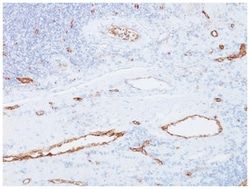
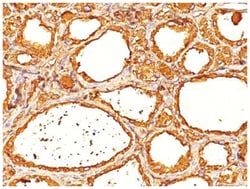
Antigen: VEGF R2/KDR/Flk-1
Dilution: Flow Cytometry 0.5 - 1ug/million cells in 0.1ml, Immunofluorescence 0.5 - 1ug/ml
Classification: Monoclonal
Form: Purified
Regulatory Status: RUO
Target Species: Human
Gene Accession No.: P35968
Gene ID (Entrez): 3791
Immunogen: Recombinant human VEGF-R2 protein
Primary or Secondary: Primary
Content And Storage: Store at 4C.
Clone: KDR657
Applications: Flow Cytometry, Immunofluorescence
Conjugate: Unconjugated
Host Species: Mouse
Research Discipline: Angiogenesis, Cancer, Cellular Markers, Endothelial Cell Markers, Hematopoietic Stem Cell Markers, HIF Target Genes, Hypoxia, Phospho Specific, Signal Transduction, Stem Cell Markers, Tumor Suppressors, Tyrosine Kinases
Formulation: PBS with 0.05% BSA. with 0.05% Sodium Azide
Gene Alias: CD309, CD309 antigen, EC 2.7.10, EC 2.7.10.1, Fetal liver kinase 1, fetal liver kinase-1, FLK-1, FLK1tyrosine kinase growth factor receptor, Kinase insert domain receptor, kinase insert domain receptor (a type III receptor tyrosine kinase), Protein-tyrosine kinase receptor flk-1, soluble VEGFR2, vascular endothelial growth factor receptor 2, VEGFR, VEGFR2, VEGFR-2
Gene Symbols: KDR
Isotype: IgG1 κ
Purification Method: Protein A purified
Test Specificity: CD309, also known as VEGF-R2, KDR3, and Flk-1 (mouse), is a type I transmembrane glycoprotein. It is a member of the CSF-1/PDGF receptor family of type III tyrosine kinase receptors. Human VEGF-R2 is mainly expressed by endothelial cells, embryonic tissues, and megakaryocytes. It plays an important role in the regulation of angiogenesis, vasculogenesis, and vascular permeability. The ligands of VEGF-R2 include VEGF-A, VEGF-C, VEGF-D, and VEGF splice isoforms. Ligation of VEGF-R2 with its ligands results in the receptor dimerization and auto-phosphorylation, stimulating endothelial cell proliferation and migration.
Antigen: VEGF R2/KDR/Flk-1
Dilution: Flow Cytometry 0.5-1ug/million cells, Immunocytochemistry/Immunofluorescence 0.5-1ug/ml, Immunoprecipitation 0.5-1ug/500ug protein lysate, Immunohistochemistry-Frozen 0.5-1.0ug/ml
Classification: Monoclonal
Form: Purified
Regulatory Status: RUO
Target Species: Human
Gene Accession No.: P35968
Gene ID (Entrez): 3791
Immunogen: Recombinant human VEGF-R2 protein
Primary or Secondary: Primary
Content And Storage: Store at 4C.
Clone: KDR721
Applications: Flow Cytometry, Immunocytochemistry, Immunofluorescence, Immunoprecipitation, Immunohistochemistry (Frozen)
Conjugate: Unconjugated
Host Species: Mouse
Research Discipline: Angiogenesis, Cancer, Cellular Markers, Endothelial Cell Markers, Hematopoietic Stem Cell Markers, HIF Target Genes, Hypoxia, Phospho Specific, Signal Transduction, Stem Cell Markers, Tumor Suppressors, Tyrosine Kinases
Formulation: PBS with 0.05% BSA. with 0.05% Sodium Azide
Gene Alias: CD309, CD309 antigen, EC 2.7.10, EC 2.7.10.1, Fetal liver kinase 1, fetal liver kinase-1, FLK-1, FLK1tyrosine kinase growth factor receptor, Kinase insert domain receptor, kinase insert domain receptor (a type III receptor tyrosine kinase), Protein-tyrosine kinase receptor flk-1, soluble VEGFR2, vascular endothelial growth factor receptor 2, VEGFR, VEGFR2, VEGFR-2
Gene Symbols: KDR
Isotype: IgG1 κ
Purification Method: Protein A purified
Test Specificity: CD309, also known as VEGFR2, KDR3, and Flk-1 (mouse), is a type I transmembrane glycoprotein. It is a member of the CSF-1/PDGF receptor family of type III tyrosine kinase receptors. Human VEGFR2 is mainly expressed by endothelial cells, embryonic tissues, and megakaryocytes. It plays an important role in the regulation of angiogenesis, vasculogenesis, and vascular permeability. The ligands of VEGFR2 include VEGF-A, VEGF-C, VEGF-D, and VEGF splice isoforms. Ligation of VEGFR2 with its ligands results in the receptor dimerization and auto-phosphorylation, stimulating endothelial cell proliferation and migration.