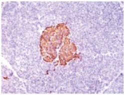
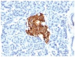
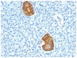
Insulin Antibody (IRDN/805) - Azide and BSA Free, Novus Biologicals™
Mouse Monoclonal Antibody
Manufacturer: Fischer Scientific
The price for this product is unavailable. Please request a quote
Antigen
Insulin
Concentration
1.0 mg/mL
Applications
Flow Cytometry, Immunohistochemistry (Paraffin), Immunofluorescence, CyTOF
Conjugate
Unconjugated
Host Species
Mouse
Research Discipline
Diabetes Research, Immune System Diseases, Immunology, Stem Cell Markers
Formulation
PBS with No Preservative
Gene ID (Entrez)
3630
Immunogen
Recombinant INS protein
Primary or Secondary
Primary
Content And Storage
Store at 4C short term. Aliquot and store at -20C long term. Avoid freeze-thaw cycles.
Molecular Weight of Antigen
6 kDa
Clone
IRDN/805
Dilution
Flow Cytometry : 0.5 - 1 ug/million cells in 0.1 ml, Immunohistochemistry-Paraffin : 0.5 - 1.0 ug/ml, Immunofluorescence : 1 - 2 ug/ml, CyTOF-ready
Classification
Monoclonal
Form
Purified
Regulatory Status
RUO
Target Species
Human, Mouse, Porcine, Primate, Rabbit, Rat (Negative)
Gene Alias
IDDM2, ILPR, insulin, IRDN, MODY10, proinsulin
Gene Symbols
INS
Isotype
IgG1 κ
Purification Method
Protein A or G purified
Test Specificity
Recognizes a polypeptide which is identified as insulin, a 51-amino acid polypeptide composed of A and B chains connected through the C-peptide. Proinsulin, which has very little biological activity, is cleaved by proteases within its cell of origin into the insulin molecule and the C-terminal basic residue. Insulin enhances membrane transport of glucose, amino acids, and certain ions. It also promotes glycogen storage, formation of triglycerides, and synthesis of proteins and nucleic acids. Deficiency of insulin results in diabetes mellitus. The main storage site for insulin is the pancreatic islets. Antibodies to insulin are important as beta-cell and insulinoma marker.
Related Products

Description
- Insulin Monoclonal specifically detects Insulin in Human, Mouse, Porcine, Bovine, Rabbit, Rat (Negative) samples
- It is validated for Immunohistochemistry, Immunohistochemistry-Paraffin.
Compare Similar Items
Show Difference
Antigen: Insulin
Concentration: 1.0 mg/mL
Applications: Flow Cytometry, Immunohistochemistry (Paraffin), Immunofluorescence, CyTOF
Conjugate: Unconjugated
Host Species: Mouse
Research Discipline: Diabetes Research, Immune System Diseases, Immunology, Stem Cell Markers
Formulation: PBS with No Preservative
Gene ID (Entrez): 3630
Immunogen: Recombinant INS protein
Primary or Secondary: Primary
Content And Storage: Store at 4C short term. Aliquot and store at -20C long term. Avoid freeze-thaw cycles.
Molecular Weight of Antigen: 6 kDa
Clone: IRDN/805
Dilution: Flow Cytometry : 0.5 - 1 ug/million cells in 0.1 ml, Immunohistochemistry-Paraffin : 0.5 - 1.0 ug/ml, Immunofluorescence : 1 - 2 ug/ml, CyTOF-ready
Classification: Monoclonal
Form: Purified
Regulatory Status: RUO
Target Species: Human, Mouse, Porcine, Primate, Rabbit, Rat (Negative)
Gene Alias: IDDM2, ILPR, insulin, IRDN, MODY10, proinsulin
Gene Symbols: INS
Isotype: IgG1 κ
Purification Method: Protein A or G purified
Test Specificity: Recognizes a polypeptide which is identified as insulin, a 51-amino acid polypeptide composed of A and B chains connected through the C-peptide. Proinsulin, which has very little biological activity, is cleaved by proteases within its cell of origin into the insulin molecule and the C-terminal basic residue. Insulin enhances membrane transport of glucose, amino acids, and certain ions. It also promotes glycogen storage, formation of triglycerides, and synthesis of proteins and nucleic acids. Deficiency of insulin results in diabetes mellitus. The main storage site for insulin is the pancreatic islets. Antibodies to insulin are important as beta-cell and insulinoma marker.
Antigen: PTH
Concentration: 1.0 mg/mL
Applications: Flow Cytometry, Immunohistochemistry (Paraffin), Immunofluorescence, CyTOF
Conjugate: Unconjugated
Host Species: Mouse
Research Discipline: Apoptosis, Cancer
Formulation: PBS with No Preservative
Gene ID (Entrez): 5741
Immunogen: Recombinant fragment (84 amino acid residues from C-terminus) of human PTH protein
Primary or Secondary: Primary
Content And Storage: Store at 4C short term. Aliquot and store at -20C long term. Avoid freeze-thaw cycles.
Molecular Weight of Antigen: 9 kDa
Clone: PTH/1174
Dilution: Flow Cytometry : 0.5 - 1 ug/million cells in 0.1 ml, Immunohistochemistry-Paraffin : 0.5 - 1.0 ug/ml, Immunofluorescence : 0.5 - 1.0 ug/ml, CyTOF-ready
Classification: Monoclonal
Form: Purified
Regulatory Status: RUO
Target Species: Human
Gene Alias: Parathormone, Parathyrin, parathyroid hormone, parathyroid hormone 1, PTH1
Gene Symbols: PTH
Isotype: IgG2b κ
Purification Method: Protein A or G purified
Test Specificity: Epitope of this MAb maps in the C-terminus of PTH, a hormone produced by the parathyroid gland that regulates the concentration of calcium and phosphorus in extracellular fluid. This hormone elevates blood Ca2+ levels by dissolving the salts in bone and preventing their renal excretion.It is produced in the parathyroid gland as an 84 amino acid single chain polypeptide. It can also be secreted as N-terminal truncated fragments or C-terminal fragments after intracellular degradation, as in case of hypercalcemia. Defects in this gene are a cause of familial isolated hypoparathyroidism (FIH); also called autosomal dominant hypoparathyroidism or autosomal dominant hypocalcemia. FIH is characterized by hypocalcemia and hyperphosphatemia due to inadequate secretion of parathyroid hormone. Symptoms are seizures, tetany and cramps. FIH exist both as autosomal dominant and recessive forms of hypoparathyroidism.
Antigen: LH beta
Concentration: 1.0 mg/mL
Applications: Flow Cytometry, Immunohistochemistry (Paraffin), Immunofluorescence, CyTOF
Conjugate: Unconjugated
Host Species: Mouse
Research Discipline: __
Formulation: PBS with No Preservative
Gene ID (Entrez): 3972
Immunogen: Recombinant beta sub-unit of human LH
Primary or Secondary: Primary
Content And Storage: Store at 4C short term. Aliquot and store at -20C long term. Avoid freeze-thaw cycles.
Molecular Weight of Antigen: 22 kDa
Clone: LHb/1214
Dilution: Flow Cytometry : 0.5 - 1 ug/million cells in 0.1 ml, Immunohistochemistry-Paraffin : 0.5 - 1.0 ug/ml, Immunofluorescence : 1 - 2 ug/ml, CyTOF-ready
Classification: Monoclonal
Form: Purified
Regulatory Status: RUO
Target Species: Human
Gene Alias: CGB4, hLHB, interstitial cell stimulating hormone, beta chain, LSH-B, LSH-beta, luteinizing hormone beta polypeptide, luteinizing hormone beta subunit, lutropin beta chain, lutropin subunit beta
Gene Symbols: LHB
Isotype: IgG1 κ
Purification Method: Protein A or G purified
Test Specificity: Luteinizing hormone (LH) is a glycoprotein. Each monomeric unit is a sugar-like protein molecule; two of these make the full, functional protein. Its structure is similar to the other glycoproteins, follicle-stimulating hormone (FSH), thyroid-stimulating hormone (TSH), and human chorionic gonadotropin (hCG). The protein dimer contains 2 polypeptide units, labeled alpha and beta subunits that are connected by two bridges. The alpha subunits of LH, FSH, TSH, and hCG are identical, and contain 92 amino acids. The beta subunits vary. LH has a beta subunit of 121 amino acids (LHB) that confers its specific biologic action and is responsible for interaction with the LH receptor. This beta subunit contains the same amino acids in sequence as the beta subunit of hCG and both stimulate the same receptor; however, the hCG beta subunit contains an additional 24 amino acids and the hormones differ in the composition of their sugar moieties.LH is synthesized and secreted by gonadotrophs in the anterior lobe of the pituitary gland. In concert with the other pituitary gonadotropin follicle-stimulating hormone (FSH), it is necessary for proper reproductive function. In the female, an acute rise of LH levels triggers ovulation. In the male, where LH has also been called Interstitial Cell-Stimulating Hormone (ICSH), it stimulates Leydig cell production of testosterone. LH is a useful marker in classification of pituitary tumors and the study of pituitary disease.