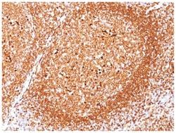
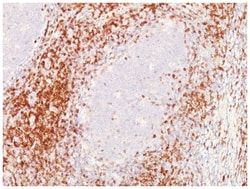
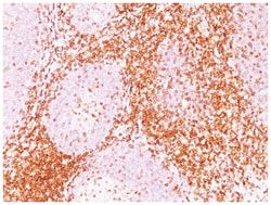
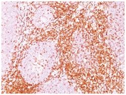
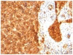
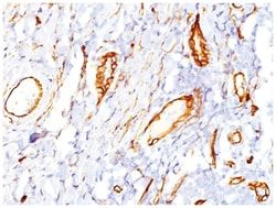
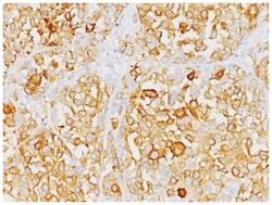
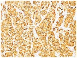
CD63 Antibody (SPM524), Novus Biologicals™
Mouse Monoclonal Antibody has been used in 1 publication
Manufacturer: Fischer Scientific
The price for this product is unavailable. Please request a quote
Antigen
CD63
Dilution
Western Blot 0.5-1ug/ml, Flow Cytometry 0.5-1ug/million cells, Immunocytochemistry/Immunofluorescence 0.5-1ug/ml, Immunoprecipitation 0.5-1ug/500ug protein lysate, Immunohistochemistry-Paraffin 0.5-1ug/ml, Immunohistochemistry-Frozen 0.5-1.0ug/ml
Classification
Monoclonal
Form
Purified
Regulatory Status
RUO
Target Species
Human, Mouse
Gene Accession No.
P08962
Gene ID (Entrez)
967
Immunogen
Recombinant human CD63 protein
Primary or Secondary
Primary
Content And Storage
Store at 4C.
Clone
SPM524
Applications
Western Blot, Flow Cytometry, Immunocytochemistry, Immunofluorescence, Immunoprecipitation, Immunohistochemistry (Paraffin)
Conjugate
Unconjugated
Host Species
Mouse
Research Discipline
Autophagy, Cytokine Research
Formulation
PBS with 0.05% BSA. with 0.05% Sodium Azide
Gene Alias
CD63 antigen, CD63 antigen (melanoma 1 antigen), CD63 molecule, Granulophysin, LAMP-3, Lysosomal-associated membrane protein 3, ME491, melanoma 1 antigen, Melanoma-associated antigen ME491, melanoma-associated antigen MLA1, MLA1lysosome-associated membrane glycoprotein 3, Ocular melanoma-associated antigen, OMA81H, Tetraspanin-30, tspan-30, TSPAN30granulophysin
Gene Symbols
CD63
Isotype
IgG1 κ
Purification Method
Protein A purified
Test Specificity
This product is specific for CD63
Related Products
Description
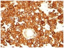
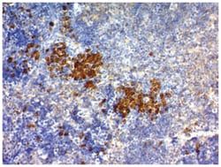
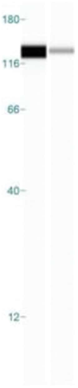
- CD63 Monoclonal specifically detects CD63 in Human, Mouse samples
- It is validated for Western Blot, Flow Cytometry, Immunohistochemistry, Immunocytochemistry/Immunofluorescence, Immunohistochemistry-Paraffin.
Compare Similar Items
Show Difference
Antigen: CD63
Dilution: Western Blot 0.5-1ug/ml, Flow Cytometry 0.5-1ug/million cells, Immunocytochemistry/Immunofluorescence 0.5-1ug/ml, Immunoprecipitation 0.5-1ug/500ug protein lysate, Immunohistochemistry-Paraffin 0.5-1ug/ml, Immunohistochemistry-Frozen 0.5-1.0ug/ml
Classification: Monoclonal
Form: Purified
Regulatory Status: RUO
Target Species: Human, Mouse
Gene Accession No.: P08962
Gene ID (Entrez): 967
Immunogen: Recombinant human CD63 protein
Primary or Secondary: Primary
Content And Storage: Store at 4C.
Clone: SPM524
Applications: Western Blot, Flow Cytometry, Immunocytochemistry, Immunofluorescence, Immunoprecipitation, Immunohistochemistry (Paraffin)
Conjugate: Unconjugated
Host Species: Mouse
Research Discipline: Autophagy, Cytokine Research
Formulation: PBS with 0.05% BSA. with 0.05% Sodium Azide
Gene Alias: CD63 antigen, CD63 antigen (melanoma 1 antigen), CD63 molecule, Granulophysin, LAMP-3, Lysosomal-associated membrane protein 3, ME491, melanoma 1 antigen, Melanoma-associated antigen ME491, melanoma-associated antigen MLA1, MLA1lysosome-associated membrane glycoprotein 3, Ocular melanoma-associated antigen, OMA81H, Tetraspanin-30, tspan-30, TSPAN30granulophysin
Gene Symbols: CD63
Isotype: IgG1 κ
Purification Method: Protein A purified
Test Specificity: This product is specific for CD63
Antigen: TfR (Transferrin R)
Dilution: Western Blot 0.25-0.5ug/ml, Simple Western 10 ug/ml, Flow Cytometry 0.5-1ug/million cells, Immunocytochemistry/Immunofluorescence 1-2ug/ml, Immunohistochemistry-Paraffin 0.5-1ug/ml, Immunohistochemistry-Frozen 0.5-1.0ug/ml
Classification: Monoclonal
Form: Purified
Regulatory Status: RUO
Target Species: Human
Gene Accession No.: P02786
Gene ID (Entrez): 7037
Immunogen: KG1 acute myeloid leukemia cell line
Primary or Secondary: Primary
Content And Storage: Store at 4C.
Clone: DF1513
Applications: Western Blot, Flow Cytometry, Immunocytochemistry, Immunofluorescence, Immunohistochemistry (Paraffin)
Conjugate: Unconjugated
Host Species: Mouse
Research Discipline: Cell Biology, Cellular Markers, Hematopoietic Stem Cell Markers, Immunology, Signal Transduction, Stem Cell Markers
Formulation: PBS with 0.05% BSA. with 0.05% Sodium Azide
Gene Alias: CD71 antigen, CD71TRFR, p90, T9, TfR, TFR1, TR, transferrin receptor (p90, CD71), transferrin receptor protein 1, Trfr
Gene Symbols: TFRC
Isotype: IgG1 κ
Purification Method: Protein A purified
Test Specificity: It recognizes a ∼90-95kDa protein which is identified as cell surface transferrin receptor (CD71), a disulfide-bonded homodimeric glycoprotein of 180-190kDa (Workshop IV). This MAb is highly specific to CD71 and shows no cross-reaction with other related proteins. Ligand for transferrin receptor is the serum iron transport protein, transferrin. This receptor is broadly distributed in carcinomas, sarcomas, leukemias, and lymphomas. CD71/Transferrin receptor has been reported to be associated with cell proliferation in both normal and neoplastic tissues and useful in predicting clinical behavior or response to therapy in a number of malignancies including breast cancer.

Antigen: CD74
Dilution: Western Blot 0.5-1ug/ml, Flow Cytometry 0.5-1ug/million cells, Immunocytochemistry/Immunofluorescence 1-2ug/ml, Immunoprecipitation 0.5-1ug/500ug protein lysate, Immunohistochemistry-Frozen 0.5-1ug/ml
Classification: Monoclonal
Form: Purified
Regulatory Status: RUO
Target Species: Human
Gene Accession No.: P044233
Gene ID (Entrez): 972
Immunogen: B lymphoblastoid cell line HFB1
Primary or Secondary: Primary
Content And Storage: Store at 4C.
Clone: BU45
Applications: Western Blot, Flow Cytometry, Immunocytochemistry, Immunofluorescence, Immunoprecipitation, Immunohistochemistry (Frozen)
Conjugate: Unconjugated
Host Species: Mouse
Research Discipline: Immunology
Formulation: PBS with 0.05% BSA. with 0.05% Sodium Azide
Gene Alias: CD74 antigen, CD74 antigen (invariant polypeptide of major histocompatibility complex, classII antigen-associated), CD74 molecule, major histocompatibility complex, class II invariant chain, DHLAGgamma chain of class II antigens, HLA class II histocompatibility antigen gamma chain, HLADG, HLA-DR antigens-associated invariant chain, HLA-DR-gamma, Ia antigen-associated invariant chain, Ia-associated invariant chain, Ia-GAMMA, Ii, MHC HLA-DR gamma chain, p33
Gene Symbols: CD74
Isotype: IgG1 κ
Purification Method: Protein A purified
Test Specificity: Recognizes proteins of 33, 35 and 41kDa, which are identified as various isoforms of CD74. Its epitope is localized in the extracellular domain of CD74. CD74 is a type II transmembrane protein which binds to the peptide binding groove of newly synthesized MHC class II alpha/beta heterodimers and prevents their premature association with endogenous polypeptides. The CD74 molecule plays a critical role in the presentation of peptides, by the MHC class II antigens, to CD4 positive lymphocytes. CD74 is expressed on MHC class II positive cells including B cells, a subset of activated T cells, monocytes, and dendritic cells and by various types of carcinomas. CD74 is expressed primarily by antigen presenting cells, such as B-lymphocytes (from before the pre-B cell stage to before the plasma cell stage), macrophages, and monocytes, and many epithelial cells. Anti-CD74 stains predominantly germinal center lymphocytes and B-cell lymphomas, but rarely T-cell lymphomas. Anti-CD74 has been shown to be useful in differentiating atypical fibroxanthoma (-) from malignant fibrous histiocytoma (+).