Integrin beta 4/CD104 Antibody (UM-A9), Novus Biologicals™
Mouse Monoclonal Antibody
Manufacturer: Fischer Scientific
The price for this product is unavailable. Please request a quote
Antigen
Integrin beta 4/CD104
Dilution
Western Blot 0.5-1ug/ml, Flow Cytometry 0.5-1ug/million cells, Immunocytochemistry/Immunofluorescence 0.5-1ug/ml, Immunoprecipitation 0.5-1ug/500ug protein lysate, SDS-Page
Classification
Monoclonal
Form
Purified
Regulatory Status
RUO
Target Species
Human
Gene Accession No.
P16144
Gene ID (Entrez)
3691
Immunogen
Human squamous cell carcinoma (UM-SCC1)
Primary or Secondary
Primary
Content And Storage
Store at 4C.
Molecular Weight of Antigen
205 kDa
Clone
UM-A9
Applications
Western Blot, Flow Cytometry, Immunocytochemistry, Immunofluorescence, Immunoprecipitation, SDS-Page
Conjugate
Unconjugated
Host Species
Mouse
Research Discipline
Adaptive Immunity, Immunology, Phospho Specific, Signal Transduction
Formulation
PBS with 0.05% BSA. with 0.05% Sodium Azide
Gene Alias
CD104, CD104 antigen, GP150, integrin beta-4, integrin beta-4 subunit, integrin, beta 4
Gene Symbols
ITGB4
Isotype
IgG2a κ
Purification Method
Protein A purified
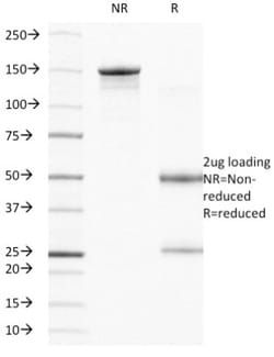
Test Specificity
Recognizes a protein of 205kDa, which is identified as integrin beta-4 (ITGB4). Its epitope is localized in the extracellular domain of ITGB4 protein. Integrins are heterodimers comprised of alpha and beta subunits, that are non-covalently associated transmembrane glycoprotein receptors. Different combinations of alpha and beta polypeptides form complexes that vary in their ligand-binding specificities. Integrins mediate cell-matrix or cell-cell adhesion, and transduced signals that regulate gene expression and cell growth. This gene encodes the integrin beta-4 subunit, a receptor for the laminins. This subunit tends to associate with alpha-6 subunit and is likely to play a pivotal role in the biology of invasive carcinoma. Mutations in this gene are associated with epidermolysis bullosa with pyloric atresia. Multiple alternatively spliced transcript variants encoding distinct isoforms have been found for this gene.
Related Products

Description
- Integrin beta 4/CD104 Monoclonal specifically detects Integrin beta 4/CD104 in Human samples
- It is validated for Flow Cytometry, Immunocytochemistry/Immunofluorescence.
Compare Similar Items
Show Difference
Antigen: Integrin beta 4/CD104
Dilution: Western Blot 0.5-1ug/ml, Flow Cytometry 0.5-1ug/million cells, Immunocytochemistry/Immunofluorescence 0.5-1ug/ml, Immunoprecipitation 0.5-1ug/500ug protein lysate, SDS-Page
Classification: Monoclonal
Form: Purified
Regulatory Status: RUO
Target Species: Human
Gene Accession No.: P16144
Gene ID (Entrez): 3691
Immunogen: Human squamous cell carcinoma (UM-SCC1)
Primary or Secondary: Primary
Content And Storage: Store at 4C.
Molecular Weight of Antigen: 205 kDa
Clone: UM-A9
Applications: Western Blot, Flow Cytometry, Immunocytochemistry, Immunofluorescence, Immunoprecipitation, SDS-Page
Conjugate: Unconjugated
Host Species: Mouse
Research Discipline: Adaptive Immunity, Immunology, Phospho Specific, Signal Transduction
Formulation: PBS with 0.05% BSA. with 0.05% Sodium Azide
Gene Alias: CD104, CD104 antigen, GP150, integrin beta-4, integrin beta-4 subunit, integrin, beta 4
Gene Symbols: ITGB4
Isotype: IgG2a κ
Purification Method: Protein A purified
Test Specificity: Recognizes a protein of 205kDa, which is identified as integrin beta-4 (ITGB4). Its epitope is localized in the extracellular domain of ITGB4 protein. Integrins are heterodimers comprised of alpha and beta subunits, that are non-covalently associated transmembrane glycoprotein receptors. Different combinations of alpha and beta polypeptides form complexes that vary in their ligand-binding specificities. Integrins mediate cell-matrix or cell-cell adhesion, and transduced signals that regulate gene expression and cell growth. This gene encodes the integrin beta-4 subunit, a receptor for the laminins. This subunit tends to associate with alpha-6 subunit and is likely to play a pivotal role in the biology of invasive carcinoma. Mutations in this gene are associated with epidermolysis bullosa with pyloric atresia. Multiple alternatively spliced transcript variants encoding distinct isoforms have been found for this gene.

Antigen: CD2
Dilution: Western Blot 0.5-1ug/ml, Flow Cytometry 0.5-1ug/million cells, Immunocytochemistry/Immunofluorescence 0.5-1ug/ml, Immunoprecipitation 0.5-1ug/500ug protein lysate
Classification: Monoclonal
Form: Purified
Regulatory Status: RUO
Target Species: Human
Gene Accession No.: P06729
Gene ID (Entrez): 914
Immunogen: Human Thymocytes/Sezary T cells
Primary or Secondary: Primary
Content And Storage: Store at 4C.
Molecular Weight of Antigen: 50 kDa
Clone: UMCD2
Applications: Western Blot, Flow Cytometry, Immunocytochemistry, Immunofluorescence, Immunoprecipitation
Conjugate: Unconjugated
Host Species: Mouse
Research Discipline: Adaptive Immunity, Apoptosis, Immunology
Formulation: PBS with 0.05% BSA. with 0.05% Sodium Azide
Gene Alias: CD2 antigen, CD2 antigen (p50), sheep red blood cell receptor, CD2 molecule, Erythrocyte receptor, FLJ46032, LFA-2, LFA-3 receptor, lymphocyte-function antigen-2, Rosette receptor, SRBC, T11, T-cell surface antigen CD2, T-cell surface antigen T11/Leu-5
Gene Symbols: CD2
Isotype: IgG2a κ
Purification Method: Protein A purified
Test Specificity: CD2 interacts through its amino-terminal domain with the extracellular domain of CD58 (also designated CD2 ligand) to mediate cell adhesion. CD2/CD58 binding can enhance antigen-specific T cell activation. CD2 is a transmembrane glycoprotein that is expressed on peripheral blood T lymphocytes, NK cells and thymocytes. CD58 is a heavily glycosylated protein with a broad tissue distribution in hematopoietic and other cells, including endothelium. Interaction between CD2 and its counter receptor LFA3 (CD58) on opposing cells optimizes immune system recognition, thereby facilitating communication between helper T lymphocytes and antigen-presenting cells, as well as between cytolytic effectors and target cells.