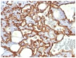
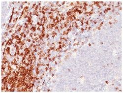
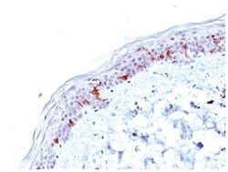
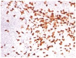
CD2 Antibody (UMCD2), Novus Biologicals™
Mouse Monoclonal Antibody
Manufacturer: Fischer Scientific
The price for this product is unavailable. Please request a quote
Antigen
CD2
Dilution
Western Blot 0.5-1ug/ml, Flow Cytometry 0.5-1ug/million cells, Immunocytochemistry/Immunofluorescence 0.5-1ug/ml, Immunoprecipitation 0.5-1ug/500ug protein lysate
Classification
Monoclonal
Form
Purified
Regulatory Status
RUO
Target Species
Human
Gene Accession No.
P06729
Gene ID (Entrez)
914
Immunogen
Human Thymocytes/Sezary T cells
Primary or Secondary
Primary
Content And Storage
Store at 4C.
Molecular Weight of Antigen
50 kDa
Clone
UMCD2
Applications
Western Blot, Flow Cytometry, Immunocytochemistry, Immunofluorescence, Immunoprecipitation
Conjugate
Unconjugated
Host Species
Mouse
Research Discipline
Adaptive Immunity, Apoptosis, Immunology
Formulation
PBS with 0.05% BSA. with 0.05% Sodium Azide
Gene Alias
CD2 antigen, CD2 antigen (p50), sheep red blood cell receptor, CD2 molecule, Erythrocyte receptor, FLJ46032, LFA-2, LFA-3 receptor, lymphocyte-function antigen-2, Rosette receptor, SRBC, T11, T-cell surface antigen CD2, T-cell surface antigen T11/Leu-5
Gene Symbols
CD2
Isotype
IgG2a κ
Purification Method
Protein A purified
Test Specificity
CD2 interacts through its amino-terminal domain with the extracellular domain of CD58 (also designated CD2 ligand) to mediate cell adhesion. CD2/CD58 binding can enhance antigen-specific T cell activation. CD2 is a transmembrane glycoprotein that is expressed on peripheral blood T lymphocytes, NK cells and thymocytes. CD58 is a heavily glycosylated protein with a broad tissue distribution in hematopoietic and other cells, including endothelium. Interaction between CD2 and its counter receptor LFA3 (CD58) on opposing cells optimizes immune system recognition, thereby facilitating communication between helper T lymphocytes and antigen-presenting cells, as well as between cytolytic effectors and target cells.
Related Products

Description
- CD2 Monoclonal specifically detects CD2 in Human samples
- It is validated for Western Blot, Flow Cytometry, Immunocytochemistry/Immunofluorescence, Functional.
Compare Similar Items
Show Difference
Antigen: CD2
Dilution: Western Blot 0.5-1ug/ml, Flow Cytometry 0.5-1ug/million cells, Immunocytochemistry/Immunofluorescence 0.5-1ug/ml, Immunoprecipitation 0.5-1ug/500ug protein lysate
Classification: Monoclonal
Form: Purified
Regulatory Status: RUO
Target Species: Human
Gene Accession No.: P06729
Gene ID (Entrez): 914
Immunogen: Human Thymocytes/Sezary T cells
Primary or Secondary: Primary
Content And Storage: Store at 4C.
Molecular Weight of Antigen: 50 kDa
Clone: UMCD2
Applications: Western Blot, Flow Cytometry, Immunocytochemistry, Immunofluorescence, Immunoprecipitation
Conjugate: Unconjugated
Host Species: Mouse
Research Discipline: Adaptive Immunity, Apoptosis, Immunology
Formulation: PBS with 0.05% BSA. with 0.05% Sodium Azide
Gene Alias: CD2 antigen, CD2 antigen (p50), sheep red blood cell receptor, CD2 molecule, Erythrocyte receptor, FLJ46032, LFA-2, LFA-3 receptor, lymphocyte-function antigen-2, Rosette receptor, SRBC, T11, T-cell surface antigen CD2, T-cell surface antigen T11/Leu-5
Gene Symbols: CD2
Isotype: IgG2a κ
Purification Method: Protein A purified
Test Specificity: CD2 interacts through its amino-terminal domain with the extracellular domain of CD58 (also designated CD2 ligand) to mediate cell adhesion. CD2/CD58 binding can enhance antigen-specific T cell activation. CD2 is a transmembrane glycoprotein that is expressed on peripheral blood T lymphocytes, NK cells and thymocytes. CD58 is a heavily glycosylated protein with a broad tissue distribution in hematopoietic and other cells, including endothelium. Interaction between CD2 and its counter receptor LFA3 (CD58) on opposing cells optimizes immune system recognition, thereby facilitating communication between helper T lymphocytes and antigen-presenting cells, as well as between cytolytic effectors and target cells.
Antigen: CD28
Dilution: Flow Cytometry 0.5-1ug/million cells in 0.1ml, Immunocytochemistry/Immunofluorescence 0.5-1ug/ml, Immunohistochemistry-Paraffin 0.5-1.0ug/ml
Classification: Monoclonal
Form: Purified
Regulatory Status: RUO
Target Species: Human
Gene Accession No.: P10747
Gene ID (Entrez): 940
Immunogen: Recombinant human CD28 protein
Primary or Secondary: Primary
Content And Storage: Store at 4C.
Molecular Weight of Antigen: __
Clone: C28/75
Applications: Flow Cytometry, Immunocytochemistry, Immunofluorescence, Immunohistochemistry (Paraffin)
Conjugate: Unconjugated
Host Species: Mouse
Research Discipline: Adaptive Immunity, Apoptosis, Immunology
Formulation: PBS with 0.05% BSA. with 0.05% Sodium Azide
Gene Alias: CD28 antigen, CD28 antigen (Tp44), CD28 molecule, MGC138290, T-cell-specific surface glycoprotein CD28, Tp44
Gene Symbols: CD28
Isotype: IgG1 κ
Purification Method: Protein A purified
Test Specificity: Recognizes a glycoprotein of 44-88kDa, which is identified as CD28. It is the critical T-cell co-stimulatory receptor which provides to the cell the important second activation signal by binding CD80 and CD86 that are expressed by antigen presenting cells. Besides its co-stimulation role, CD28 functions in preventing T-cells from anergic hyporesponsive state or from undergoing premature apoptotic cell death. CD28 is also expressed on human fetal NK cells and some NK cell lines, whereas on murine NK cells the CD28 expression is much broader.
Antigen: CD28
Dilution: Flow Cytometry 0.5-1ug/million cells, Immunocytochemistry/Immunofluorescence 0.5-1ug/ml, Immunohistochemistry-Paraffin 0.5-1.0ug/ml, Immunohistochemistry-Frozen 0.5-1.0ug/ml
Classification: Monoclonal
Form: Purified
Regulatory Status: RUO
Target Species: Human
Gene Accession No.: P10747
Gene ID (Entrez): 940
Immunogen: Recombinant human CD28 protein
Primary or Secondary: Primary
Content And Storage: Store at 4C.
Molecular Weight of Antigen: __
Clone: C28/77
Applications: Flow Cytometry, Immunocytochemistry, Immunofluorescence, Immunohistochemistry (Paraffin), Immunohistochemistry (Frozen)
Conjugate: Unconjugated
Host Species: Mouse
Research Discipline: Adaptive Immunity, Apoptosis, Immunology
Formulation: PBS with 0.05% BSA. with 0.05% Sodium Azide
Gene Alias: CD28 antigen, CD28 antigen (Tp44), CD28 molecule, MGC138290, T-cell-specific surface glycoprotein CD28, Tp44
Gene Symbols: CD28
Isotype: IgG1 κ
Purification Method: Protein A purified
Test Specificity: Recognizes a glycoprotein of 44-88kDa, which is identified as CD28. It is the critical T-cell co-stimulatory receptor which provides to the cell the important second activation signal by binding CD80 and CD86 that are expressed by antigen presenting cells. Besides its co-stimulation role, CD28 functions in preventing T-cells from anergic hyporesponsive state or from undergoing premature apoptotic cell death. CD28 is also expressed on human fetal NK cells and some NK cell lines, whereas on murine NK cells the CD28 expression is much broader.