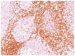
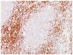
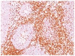
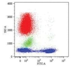
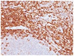
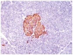
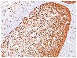
CD28 Antibody (C28/77), Novus Biologicals™
Mouse Monoclonal Antibody has been used in 1 publication
Manufacturer: Fischer Scientific
The price for this product is unavailable. Please request a quote
Antigen
CD28
Dilution
Flow Cytometry 0.5-1ug/million cells, Immunocytochemistry/Immunofluorescence 0.5-1ug/ml, Immunohistochemistry-Paraffin 0.5-1.0ug/ml, Immunohistochemistry-Frozen 0.5-1.0ug/ml
Classification
Monoclonal
Form
Purified
Regulatory Status
RUO
Target Species
Human
Gene Accession No.
P10747
Gene ID (Entrez)
940
Immunogen
Recombinant human CD28 protein
Primary or Secondary
Primary
Content And Storage
Store at 4C.
Clone
C28/77
Applications
Flow Cytometry, Immunocytochemistry, Immunofluorescence, Immunohistochemistry (Paraffin), Immunohistochemistry (Frozen)
Conjugate
Unconjugated
Host Species
Mouse
Research Discipline
Adaptive Immunity, Apoptosis, Immunology
Formulation
PBS with 0.05% BSA. with 0.05% Sodium Azide
Gene Alias
CD28 antigen, CD28 antigen (Tp44), CD28 molecule, MGC138290, T-cell-specific surface glycoprotein CD28, Tp44
Gene Symbols
CD28
Isotype
IgG1 κ
Purification Method
Protein A purified
Test Specificity
Recognizes a glycoprotein of 44-88kDa, which is identified as CD28. It is the critical T-cell co-stimulatory receptor which provides to the cell the important second activation signal by binding CD80 and CD86 that are expressed by antigen presenting cells. Besides its co-stimulation role, CD28 functions in preventing T-cells from anergic hyporesponsive state or from undergoing premature apoptotic cell death. CD28 is also expressed on human fetal NK cells and some NK cell lines, whereas on murine NK cells the CD28 expression is much broader.
Related Products

Description
- CD28 Monoclonal specifically detects CD28 in Human samples
- It is validated for ELISA.
Compare Similar Items
Show Difference
Antigen: CD28
Dilution: Flow Cytometry 0.5-1ug/million cells, Immunocytochemistry/Immunofluorescence 0.5-1ug/ml, Immunohistochemistry-Paraffin 0.5-1.0ug/ml, Immunohistochemistry-Frozen 0.5-1.0ug/ml
Classification: Monoclonal
Form: Purified
Regulatory Status: RUO
Target Species: Human
Gene Accession No.: P10747
Gene ID (Entrez): 940
Immunogen: Recombinant human CD28 protein
Primary or Secondary: Primary
Content And Storage: Store at 4C.
Clone: C28/77
Applications: Flow Cytometry, Immunocytochemistry, Immunofluorescence, Immunohistochemistry (Paraffin), Immunohistochemistry (Frozen)
Conjugate: Unconjugated
Host Species: Mouse
Research Discipline: Adaptive Immunity, Apoptosis, Immunology
Formulation: PBS with 0.05% BSA. with 0.05% Sodium Azide
Gene Alias: CD28 antigen, CD28 antigen (Tp44), CD28 molecule, MGC138290, T-cell-specific surface glycoprotein CD28, Tp44
Gene Symbols: CD28
Isotype: IgG1 κ
Purification Method: Protein A purified
Test Specificity: Recognizes a glycoprotein of 44-88kDa, which is identified as CD28. It is the critical T-cell co-stimulatory receptor which provides to the cell the important second activation signal by binding CD80 and CD86 that are expressed by antigen presenting cells. Besides its co-stimulation role, CD28 functions in preventing T-cells from anergic hyporesponsive state or from undergoing premature apoptotic cell death. CD28 is also expressed on human fetal NK cells and some NK cell lines, whereas on murine NK cells the CD28 expression is much broader.
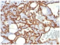
Antigen: CD31/PECAM-1
Dilution: Flow Cytometry 0.5-1ug/million cells, Immunocytochemistry/Immunofluorescence 1-2ug/ml, Immunohistochemistry-Paraffin 0.5-1.0ug/ml
Classification: Monoclonal
Form: Purified
Regulatory Status: RUO
Target Species: Human
Gene Accession No.: P16284
Gene ID (Entrez): 5175
Immunogen: Human recombinant CD31 protein
Primary or Secondary: Primary
Content And Storage: Store at 4C.
Clone: SPM532
Applications: Flow Cytometry, Immunocytochemistry, Immunofluorescence, Immunohistochemistry (Paraffin)
Conjugate: Unconjugated
Host Species: Mouse
Research Discipline: Angiogenesis, Cancer, Cellular Markers, Cytoskeleton Markers, Embryonic Stem Cell Markers, Endothelial Cell Markers, Extracellular Matrix, Hematopoietic Stem Cell Markers, Immunology, Mesenchymal Stem Cell Markers, Myeloid Cell Markers, Signal Transduction, Stem Cell Markers
Formulation: 10mM PBS and 0.05% BSA with 0.05% Sodium Azide
Gene Alias: adhesion molecule, CD31, CD31 antigen, CD31/EndoCAM, EndoCAM, FLJ34100, FLJ58394, GPIIA', PECA1, PECAM-1, PECAM-1, CD31/EndoCAM, platelet endothelial cell adhesion molecule, platelet/endothelial cell adhesion molecule
Gene Symbols: PECAM1
Isotype: IgG1 κ
Purification Method: Protein A or G purified
Test Specificity: CD31 (PECAM-1) is a transmembrane glycoprotein member of the immunoglobulin supergene family of adhesion molecules. CD31 is expressed by stem cells of the hematopoietic system and is primarily used to identify and concentrate these cells for experimental studies as well as for bone marrow transplantation. Anti-CD31 has shown to be highly specific and sensitive for vascular endothelial cells. Staining of nonvascular tumors (excluding hematopoietic neoplasms) is rare. CD31 MAb reacts with normal, benign, and malignant endothelial cells which make up blood vessel lining. The level of CD31 expression can help to determine the degree of tumor angiogenesis, and a high level of CD31 expression may imply a rapidly growing tumor and potentially a predictor of tumor recurrence.