TFF1/pS2 Mouse, Clone: SPM573, Novus Biologicals™
Mouse Monoclonal Antibody
Manufacturer: Fischer Scientific
The price for this product is unavailable. Please request a quote
Antigen
TFF1/pS2
Dilution
Western Blot 0.5-1.0ug/ml, Flow Cytometry 0.5-1ug/million cells, Immunocytochemistry/Immunofluorescence 0.5-1ug/ml, Immunoprecipitation 0.5-1ug/500ug protein lysate, Immunohistochemistry-Paraffin, Immunohistochemistry-Frozen 0.5-1.0ug/ml
Classification
Monoclonal
Form
Purified
Regulatory Status
RUO
Target Species
Human, Primate
Gene Accession No.
P04155
Gene ID (Entrez)
7031
Immunogen
Synthetic peptide of 28 amino acid residues corresponding to CFDDTVRGVPWCFYPNTIDVPPEEECEF (aa57-84) from the C-terminus of human pS2.
Primary or Secondary
Primary
Content And Storage
Store at 4C.
Molecular Weight of Antigen
6.5 kDa
Clone
SPM573
Applications
Western Blot, Flow Cytometry, Immunocytochemistry, Immunofluorescence, Immunoprecipitation, Immunohistochemistry (Paraffin)
Conjugate
Unconjugated
Host Species
Mouse
Research Discipline
Breast Cancer, Cancer
Formulation
PBS with 0.05% BSA. with 0.05% Sodium Azide
Gene Alias
BCEIbreast cancer, estrogen-inducible sequence expressed in, Breast cancer estrogen-inducible protein, breast cancer estrogen-inducible sequence, D21S21, gastrointestinal trefoil protein pS2, hP1.A, HPS2, pNR-2, Polypeptide P1.A, Protein pS2, pS2, trefoil factor 1, trefoil factor, BCE1, human pS2 induced by estrogen from human breast cancercell line M10HP1.A
Gene Symbols
TFF1
Isotype
IgG1 κ
Purification Method
Protein A purified
Test Specificity
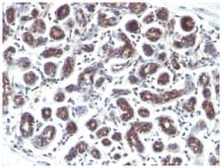
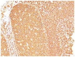
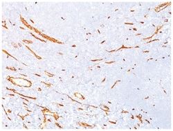
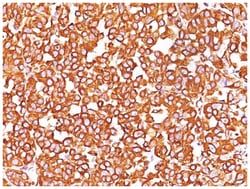
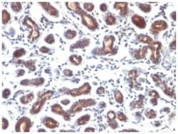
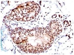
It recognizes a polypeptide of 6.5kDa, identified as pS2 estrogen-regulated protein. Its epitope is localized between aa57-84 of human pS2 protein. pS2 is a trefoil peptide. Trefoil peptides are protease resistant molecules secreted throughout the gut that play a role in mucosal healing. These peptides contain three intra-chain disulfide bonds, forming the trefoil motif, or P-domain. pS2 is known to form dimers and this dimerization is thought to play a role in its protective and healing properties. About 60% of breast carcinomas are positive for pS2. Staining is cytoplasmic, often with localization to the Golgi apparatus. pS2 is shown to be localized in normal stomach mucosa, gastric fluid, goblet cells in the colon and small intestine, and in ulcerations of the gastrointestinal tract. Several studies have shown that pS2 is primarily expressed in estrogen receptor-positive breast tumors and it may define a subset of estrogen-dependent tumors that displays an increased likelihood of re
Related Products


Description
- TFF1/pS2 Monoclonal specifically detects TFF1/pS2 in Human, Cynomolgus Monkey samples
- It is validated for Flow Cytometry, Immunohistochemistry, Immunocytochemistry/Immunofluorescence, Immunohistochemistry-Paraffin.
Compare Similar Items
Show Difference
Antigen: TFF1/pS2
Dilution: Western Blot 0.5-1.0ug/ml, Flow Cytometry 0.5-1ug/million cells, Immunocytochemistry/Immunofluorescence 0.5-1ug/ml, Immunoprecipitation 0.5-1ug/500ug protein lysate, Immunohistochemistry-Paraffin, Immunohistochemistry-Frozen 0.5-1.0ug/ml
Classification: Monoclonal
Form: Purified
Regulatory Status: RUO
Target Species: Human, Primate
Gene Accession No.: P04155
Gene ID (Entrez): 7031
Immunogen: Synthetic peptide of 28 amino acid residues corresponding to CFDDTVRGVPWCFYPNTIDVPPEEECEF (aa57-84) from the C-terminus of human pS2.
Primary or Secondary: Primary
Content And Storage: Store at 4C.
Molecular Weight of Antigen: 6.5 kDa
Clone: SPM573
Applications: Western Blot, Flow Cytometry, Immunocytochemistry, Immunofluorescence, Immunoprecipitation, Immunohistochemistry (Paraffin)
Conjugate: Unconjugated
Host Species: Mouse
Research Discipline: Breast Cancer, Cancer
Formulation: PBS with 0.05% BSA. with 0.05% Sodium Azide
Gene Alias: BCEIbreast cancer, estrogen-inducible sequence expressed in, Breast cancer estrogen-inducible protein, breast cancer estrogen-inducible sequence, D21S21, gastrointestinal trefoil protein pS2, hP1.A, HPS2, pNR-2, Polypeptide P1.A, Protein pS2, pS2, trefoil factor 1, trefoil factor, BCE1, human pS2 induced by estrogen from human breast cancercell line M10HP1.A
Gene Symbols: TFF1
Isotype: IgG1 κ
Purification Method: Protein A purified
Test Specificity: It recognizes a polypeptide of 6.5kDa, identified as pS2 estrogen-regulated protein. Its epitope is localized between aa57-84 of human pS2 protein. pS2 is a trefoil peptide. Trefoil peptides are protease resistant molecules secreted throughout the gut that play a role in mucosal healing. These peptides contain three intra-chain disulfide bonds, forming the trefoil motif, or P-domain. pS2 is known to form dimers and this dimerization is thought to play a role in its protective and healing properties. About 60% of breast carcinomas are positive for pS2. Staining is cytoplasmic, often with localization to the Golgi apparatus. pS2 is shown to be localized in normal stomach mucosa, gastric fluid, goblet cells in the colon and small intestine, and in ulcerations of the gastrointestinal tract. Several studies have shown that pS2 is primarily expressed in estrogen receptor-positive breast tumors and it may define a subset of estrogen-dependent tumors that displays an increased likelihood of re
Antigen: SUMO1
Dilution: Western Blot 0.5-1ug/ml, Flow Cytometry 0.5-1ug/million cells, Immunocytochemistry/Immunofluorescence 0.5-1ug/ml, Immunohistochemistry-Paraffin 0.5-1.0ug/ml, Immunohistochemistry-Frozen 0.5-1.0ug/ml, SDS-Page
Classification: Monoclonal
Form: Purified
Regulatory Status: RUO
Target Species: Human, Rat
Gene Accession No.: P63165
Gene ID (Entrez): 7341
Immunogen: Recombinant human SUMO1 protein
Primary or Secondary: Primary
Content And Storage: Store at 4C.
Molecular Weight of Antigen: __
Clone: SM1/495
Applications: Western Blot, Flow Cytometry, Immunocytochemistry, Immunofluorescence, Immunohistochemistry (Paraffin), Immunohistochemistry (Frozen)
Conjugate: Unconjugated
Host Species: Mouse
Research Discipline: Alzheimers Research, Core ESC Like Genes, Neuroscience, Stem Cell Markers
Formulation: PBS with 0.05% BSA. with 0.05% Sodium Azide
Gene Alias: DAP1, GAP modifying protein 1, GAP-modifying protein 1, GMP1SMT3CSMT3H3OFC10UBL1PIC1, SENP2, sentrin, small ubiquitin-related modifier 1, SMT3, SMT3 homolog 3, SMT3 suppressor of mif two 3 homolog 1 (S. cerevisiae), SMT3 suppressor of mif two 3 homolog 1 (yeast), Smt3C, SUMO-1, Ubiquitin-homology domain protein PIC1, ubiquitin-like 1 (sentrin), Ubiquitin-like protein SMT3C, Ubiquitin-like protein UBL1
Gene Symbols: SUMO1
Isotype: IgG1 κ
Purification Method: Protein A purified
Test Specificity: This MAb is specific to SUMO-1 and shows no cross-reaction with either SUMO-2 or SUMO-3. The small ubiquitin-related modifier (SUMO) proteins, which include SUMO-1, SUMO-2 and SUMO-3, belong to the ubiquitin-like protein family. Like ubiquitin, the SUMO proteins are synthesized as precursor proteins that undergo processing before conjugation to target proteins. Also, both utilize the E1, E2, and E3 cascade enzymes for conjugation. However, SUMO and ubiquitin differ with respect to targeting. Ubiquitination predominantly targets proteins for degradation, whereas sumoylation targets proteins to a variety of cellular processing, including nuclear transport, transcriptional regulation, apoptosis and protein stability. The unconjugated SUMO-1 protein localizes to the nuclear membrane.

Antigen: SUMO1
Dilution: Western Blot 0.5-1ug/ml, Flow Cytometry 0.5-1ug/million cells, Immunocytochemistry/Immunofluorescence 0.5-1ug/ml, Immunoprecipitation 0.5-1ug/500ug protein lysate, Immunohistochemistry-Paraffin 0.5-1.0ug/ml, Immunohistochemistry-Frozen 0.5-1.0ug/ml
Classification: Monoclonal
Form: Purified
Regulatory Status: RUO
Target Species: Human
Gene Accession No.: P63165
Gene ID (Entrez): 7341
Immunogen: Recombinant human SUMO1 protein
Primary or Secondary: Primary
Content And Storage: Store at 4C.
Molecular Weight of Antigen: __
Clone: SPM571
Applications: Western Blot, Flow Cytometry, Immunocytochemistry, Immunofluorescence, Immunoprecipitation, Immunohistochemistry (Paraffin)
Conjugate: Unconjugated
Host Species: Mouse
Research Discipline: Alzheimers Research, Core ESC Like Genes, Neuroscience, Stem Cell Markers
Formulation: PBS with 0.05% BSA. with 0.05% Sodium Azide
Gene Alias: DAP1, GAP modifying protein 1, GAP-modifying protein 1, GMP1SMT3CSMT3H3OFC10UBL1PIC1, SENP2, sentrin, small ubiquitin-related modifier 1, SMT3, SMT3 homolog 3, SMT3 suppressor of mif two 3 homolog 1 (S. cerevisiae), SMT3 suppressor of mif two 3 homolog 1 (yeast), Smt3C, SUMO-1, Ubiquitin-homology domain protein PIC1, ubiquitin-like 1 (sentrin), Ubiquitin-like protein SMT3C, Ubiquitin-like protein UBL1
Gene Symbols: SUMO1
Isotype: IgG1 κ
Purification Method: Protein A purified
Test Specificity: This MAb is specific to SUMO-1 and shows no cross-reaction with either SUMO-2 or SUMO-3. The small ubiquitin-related modifier (SUMO) proteins, which include SUMO-1, SUMO-2 and SUMO-3, belong to the ubiquitin-like protein family. Like ubiquitin, the SUMO proteins are synthesized as precursor proteins that undergo processing before conjugation to target proteins. Also, both utilize the E1, E2, and E3 cascade enzymes for conjugation. However, SUMO and ubiquitin differ with respect to targeting. Ubiquitination predominantly targets proteins for degradation, whereas sumoylation targets proteins to a variety of cellular processing, including nuclear transport, transcriptional regulation, apoptosis and protein stability. The unconjugated SUMO-1 protein localizes to the nuclear membrane.
